


Biliary Tract Cancer
Compliance rules
1Abstract
This guideline addresses biliary tract cancer. “Biliary tract cancer” (BTC) is an umbrella term for carcinomas of the intra- and extrahepatic (perihilar/Klatskin tumors and distal) bile ducts and the gallbladder. In international nomenclature, biliary carcinomas are also grouped under the term cholangiocarcinoma (CCA).
BTC are rare and account for less than 2% of malignant tumors in Germany.
In locally confined stages, complete surgical resection is the treatment of choice. The 5-year survival rates in these cases range from 20% to 50%, depending on the stage, patient selection, and the outcome of the resection. In locally advanced stages, treatment should generally be multimodal, particularly when the tumor is located within the liver.
In cases of technically unresectable tumors, contraindications to surgery, and in the metastatic stage, systemic therapy is the primary treatment approach. In addition to tumor-specific therapies, the treatment of tumor-related cholestasis is an essential component of multidisciplinary care and is crucial for preventing life-threatening complications (cholangiosepsis).
Carcinomas of the ampulla of Vater (papillary carcinomas) constitute a distinct group of entities, but share some similarities with distal extrahepatic biliary carcinomas and are also addressed in this guideline (see chapter 6.3.1)
2Basics
2.1Definition and Basic Information
BTC, accounting for less than 2% of all malignant tumors, are a rare and heterogeneous tumor entity [1, 2]. The nomenclature is often inconsistent. For example, the outdated term “cholangiocellular carcinoma” or the term “cholangiocarcinoma” is sometimes used as an umbrella term for all biliary carcinomas, but many authors use it only for carcinomas of the bile ducts or intrahepatic biliary carcinomas. Figure 1 provides an overview of a possible nomenclature.
The term “cholangiocellular carcinoma” is widely used but should be avoided, as the purely cholangiocellular origin of these tumors has not been sufficiently established.
2.2[Kapitel nicht relevant]
2.3Epidemiology
There are marked geographical differences in the incidence of BTC. They are rare in Europe, Australia, and the United States, with an incidence of 0.3-3.5 per 100,000 inhabitants. In countries with high rates of hepatic trematode infections, the incidence is significantly higher [3]. For extrahepatic BTC, regional differences are minimal, with an incidence ranging from 0.5 (United Kingdom) to 1.1 (Manitoba, Canada) per 100,000 inhabitants. In Western countries, there has been an increasing incidence of intrahepatic BTC in recent decades, accompanied by a decreasing incidence of extrahepatic tumors.
Gallbladder carcinomas are also rare in Western Europe and the United States, with an incidence of 1.6-2 per 100,000 inhabitants. In Chile, India, and Eastern Europe, they occur significantly more frequently, with incidences of up to 35 per 100,000. Women are affected approximately four times more often than men. According to autopsy studies, gallbladder carcinomas are considered the most common BTC [4].
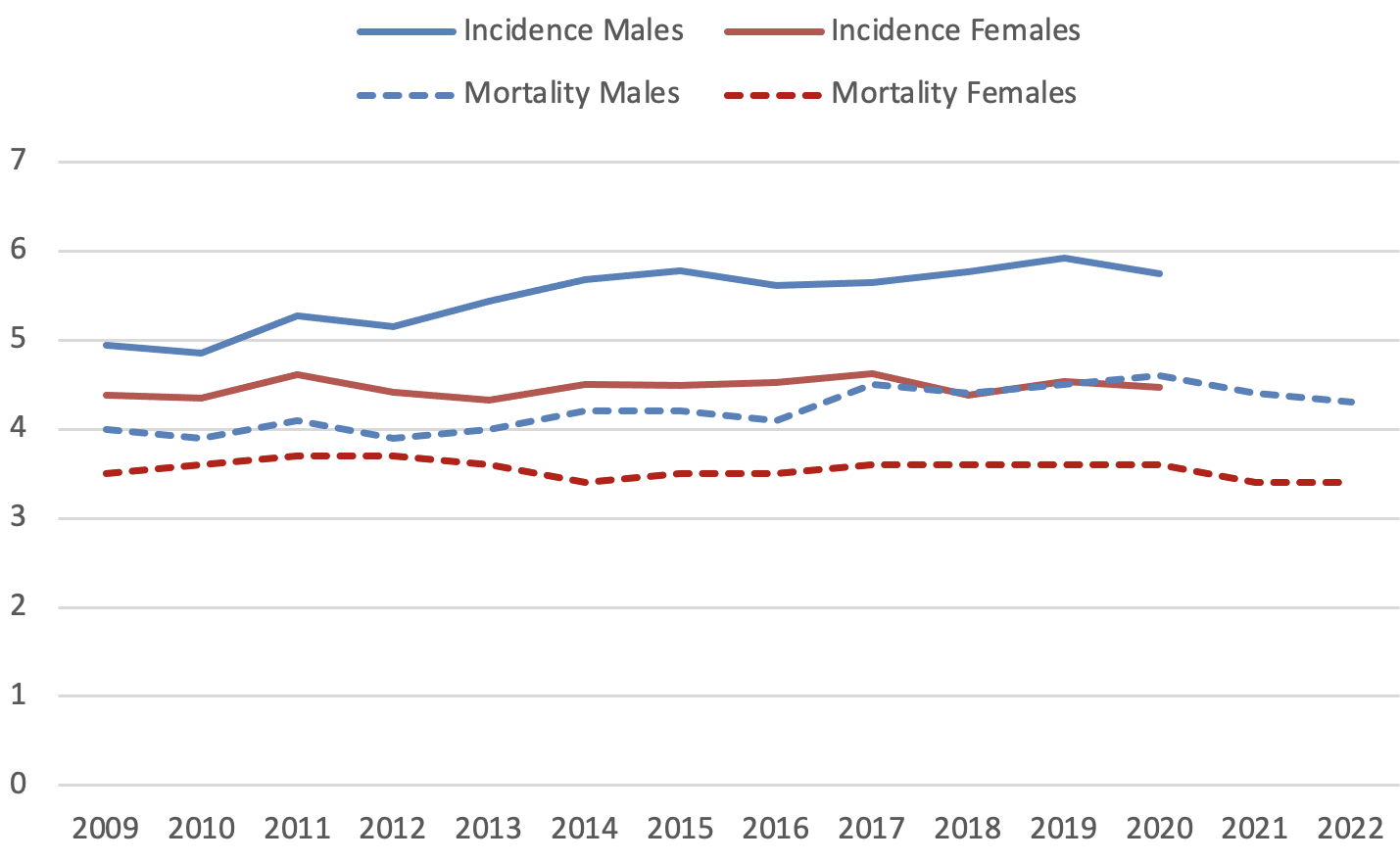
The following results are based on data from population-based cancer registries in Germany (new cases through 2024, data as of March 2026) and the nationwide statistics on causes of death from the German Federal Statistical Office.
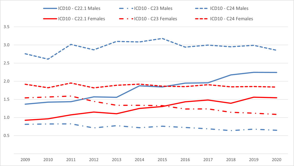
With approximately 7,700 new cases annually, BTC (malignant tumors of the gallbladder (ICD-10: C23), as well as the intra- and extrahepatic bile ducts (C22.1 and C24, respectively)) account for about 6% of all malignant tumors of the digestive tract and 1.4% of all cancers in Germany. The incidence is roughly equal between the sexes, although the gallbladder is significantly more commonly affected in women (23%) than in men (11%). Just under half of all cases (41% in women, 51% in men) involve extrahepatic BTC. Age-standardized incidence rates have remained largely constant in recent years, although the proportion of intrahepatic BTC has increased in both sexes. According to cause-of-death statistics, an average of approximately 6,500 deaths per year in Germany in recent years (2022-2024) were attributable to BTC, accounting for 2.8% of all cancer-related deaths.
The median age at diagnosis during the 2022-2024 period was 73 years for men and 75 years for women; the age distribution varies only slightly depending on tumor location.
In about one-quarter of all cases, distant metastases are already present at the time of initial diagnosis: for tumors of the gallbladder and the intrahepatic bile ducts, this proportion is around one-third, compared to extrahepatic BTCs with 15%).
The relative 5-year survival rates, which compare the observed survival to survival in the general population of the same age and sex, are currently (2020-2024) significantly lower for intrahepatic BTC with 10% compared to 21% for extrahepatic BTC.
2.4Pathogenesis
The pathogenesis of BTC is complex and also depends on the specific location. Extracellular factors such as proinflammatory cytokines, growth factors, infectious agents, or bile acids, as well as genetic aberrations causing deregulation of intracellular signaling pathways, are involved in carcinogenesis, growth, and metastasis [5]. The current WHO classification of biliary neoplasms divides intrahepatic cholangiocarcinoma into two entities, the small-duct and large-duct types, which differ morphologically, pathogenetically, and in terms of molecular pathology [6, 7]. Mutations in IDH1/2, EPHA2, or BAP1, as well as rearrangements or fusions of FGFR2, may be pathognomonic for small-duct intrahepatic cholangiocarcinomas. Mutations in ARID1A/B, PRKACA, BRAF, and RAS have been described in large-duct intrahepatic and extrahepatic cholangiocarcinomas, whereas mutations in TP53 are not location-specific and also occur very frequently in gallbladder carcinomas [8].
2.4.1Precancerous Lesions
Biliary intraepithelial neoplasia (BilIN) is considered a precursor lesion of large-duct-type intrahepatic cholangiocarcinoma, extrahepatic cholangiocarcinoma, and gallbladder carcinoma; it is divided into a low-grade and a high-grade (with a high risk of progression) type [9].
For extrahepatic cholangiocarcinoma as well as for the large-duct type of intrahepatic cholangiocarcinoma, intraductal papillary neoplasm of the biliary tract (IPNB) is also considered a preneoplasia. Based on the predominant cell type (mixed forms are common), pancreatobiliary IPNBs are distinguished from intestinal and gastric IPNBs; furthermore, they are subdivided into low-grade and high-grade forms. Depending on the specific constellation, the risk of progression varies, and in cases of associated carcinoma, the prognosis also differs. Similar to the pancreas, intraductal oncocytic neoplasms represent a distinct tumor entity with characteristic genetic alterations. Further distinguished from these lesions are intraductal tubulopapillary neoplasms (ITPN), which, analogous to the similarly named and well-defined lesions of the pancreas, also emerge in the bile ducts [10].
Other precancerous lesions of the gallbladder include intracholecystic papillary neoplasms (ICPN, various subtypes) and intracholecystic tubular neoplasms (ICTN). In ICPN lesions with a sessile growth pattern and a size greater than 1 cm, malignant transformation is likely [11, 12].
2.5Risk Factors
Risk factors vary depending on the tumor location. In general, increased age remains the primary risk factor for developing BTC in Germany.
The main risk factors for small-duct-type intrahepatic cholangiocarcinomas are similar to those for hepatocellular carcinoma, i.e., primarily liver cirrhosis and hepatitis B or C infections. For large-duct-type intrahepatic and extrahepatic cholangiocarcinomas, chronic bile duct inflammation has been identified as a risk factor, particularly primary sclerosing cholangitis and (other) bile duct strictures associated with biliary tract stones and Caroli syndrome, as well as the parasitic infections listed below in Southeast Asia.
The risk of developing bile duct carcinoma is associated with the following factors [13]:
Chronic infections, parasitic infestations (Opisthorchis viverrini, Clonorchis sinensis)
Primary sclerosing cholangitis: annual risk 0.5-1.5%, lifetime risk 5-10%
Hepatolithiasis, choledocholithiasis
Choledochal cysts
Caroli syndrome
Thorotrast as an X-ray contrast agent (no longer in use since the mid-1950s)
Additional potential risk factors for intrahepatic cholangiocarcinoma include hepatitis B and C infections, liver cirrhosis, alcohol and nicotine use, hepatic steatosis, and diabetes mellitus.
The risk of developing gallbladder cancer is increased by the following factors [14]:
Cholelithiasis (significantly increased risk for stones larger than 3 cm; for cholesterol-containing stones, possibly as small as 1.5 cm)
Porcelain gallbladder
ICPN of the gallbladder (≥ 1 cm)
Primary sclerosing cholangitis
Chronic infections, such as chronic Salmonella carriers
Biliary tract malformations (e.g., Mirizzi syndrome)
Obesity
3Prevention and Early Detection
3.1Prevention
Recommendations for the prevention of BTC focus on the acquired risk factors (see 2.5) identified to date.
3.2Early Detection
No early detection measures have been established in Germany for the asymptomatic population.
Porcelain gallbladder and Caroli syndrome, as defined precancerous conditions, are recognized indications for prophylactic cholecystectomy.
For patients with primary sclerosing cholangitis, a screening strategy involving CA 19-9 testing and MRI/MRCP every 6-12 months may be considered [15].
For patients with gallbladder polyps measuring 6 to 9 mm, at least one annual ultrasound follow-up should be performed. The “Biliary Cancer: ESMO Clinical Practice Guideline” recommends prophylactic cholecystectomy for polyps 10 mm or larger and for those that increase in size; however, no prospective study data are available on this [1].
The German AWMF S3 guideline recommends the following procedure for the treatment of gallstones [16]:
Asymptomatic patients with porcelain gallbladder: cholecystectomy
Asymptomatic patients with gallbladder stones > 3 cm in diameter: consider cholecystectomy
Patients with gallbladder stones and gallbladder polyps ≥ 1 cm: cholecystectomy regardless of symptoms.
4Clinical Characteristics
4.1Symptoms
Intrahepatic BTC, in particular, often remain asymptomatic for a long time; the following symptoms frequently do not become apparent until the tumor is locally advanced and/or metastatic:
Jaundice, cholangitis
Vomiting, nausea, loss of appetite
Upper abdominal pain, often on the right side
Palpable mass in the right upper abdomen (Courvoisier’s sign)
Weight loss, asthenia, fatigue
Ascites
Extrahepatic BTC causes bile flow obstruction at an early stage; in these cases, painless jaundice often leads to the diagnosis.
5Diagnosis
5.1[Kapitel nicht relevant]
5.2Diagnostics
5.2.1Initial Diagnosis
Contrast-enhanced imaging (MRI and/or CT) is the method of choice for (further) evaluation of a clinical suspicion and often also allows for complete staging and assessment of disease spread [1]; see Table 1.
Invasive diagnostic procedures such as endoscopic retrograde cholangiography/cholangiopancreaticography (ERC/ERCP), biopsy and brush cytology, or endoscopic ultrasound (EUS) with fine-needle aspiration cytology are reserved for cases that are not clear-cut or for patients with cholestasis and cholangitis who require decompression of the bile ducts. However, in cases of perihilar tumors without distant metastases, invasive diagnosis via ERC should only be performed after surgical evaluation and determination of the multidisciplinary - in this case, primarily surgical - treatment strategy.
In patients without significant cholestasis and with a high degree of suspicion of BTC based on imaging studies, these invasive procedures should be avoided, and the indication for surgical exploration should be applied generously.
For tumors that are in principle surgically resectable, liver function should be evaluated depending on the extent of the tumor and the patient’s preexisting medical conditions. This is mandatory for tumors located within the liver where a partial hepatectomy is planned.
In cases of inoperable BCT histological confirmation is mandatory before initiating therapy.
Table 1 provides an overview of the diagnostic procedures.
In cases of potentially resectable BTC, further evaluation of liver function using specialized techniques such as volumetry, the Limax® test, and elastography (Fibroscan®) may be necessary for surgical planning.
For intrahepatic BTC, dynamic contrast-enhanced MRI is indicated. This allows for a morphological distinction from HCC, which is characterized by early arterial contrast uptake and “washout” during the venous contrast phase.
5.3Classification
5.3.1Classification by Location
The current TNM classification (9th edition) [17] distinguishes the following based on location:
Carcinomas of the intrahepatic bile ducts (see chapter 5.3.2.1.)
Carcinomas of the perihilar bile ducts (Klatskin tumors) (see chapter 5.3.2.2.)
Carcinomas of the distal extrahepatic bile ducts (see chapter 5.3.2.3.)
Carcinomas of the gallbladder and cystic duct (see chapter 5.3.2.4.)
The classifications of carcinomas of the ampulla of Vater are addressed in chapter 6.3.1.2.
5.3.1.1Perihilar Tumors / Klatskin Tumors: Classification According to Bismuth-Corlette
This classification was developed to assess the extent of infiltration of the hepatic bifurcation and, consequently, surgical resectability. However, it refers only to the longitudinal extent of the lesion within the biliary tract system; see Table 2. This information alone is insufficient for assessing surgical resectability, which can only be evaluated in combination with contrast-enhanced cross-sectional imaging (CT/MRI).
With the currently available diagnostic methods (MRCP and ERC), the extent of the tumor is frequently underestimated, but may also be overestimated (20-30% in each case). Therefore, even a Type IV classification in cases of uncertain or borderline findings is not per se, a contraindication for exploration followed by surgical resection if indicated. Furthermore, thanks to modern surgical techniques, even Bismuth Type IV tumors can now be resected with R0 status in many cases, meaning that Bismuth Stage IV does not represent an absolute contraindication for surgery. Local contraindications most commonly result from involvement of arterial vessels.
Type | Description |
|---|---|
I | Tumor invades the common hepatic duct but not the hepatic bifurcation |
II | Tumor also invades the hepatic bifurcation |
IIIa IIIb | Tumor invades the hepatic bifurcation as well as the right main branch |
Tumor invades the hepatic bifurcation and the left main branch | |
IV | Tumor invades the hepatic bifurcation and both main branches |
5.3.1.2Perihilar Tumors/Klatskin Tumors: Jarnagin-Blumgart/MSK Classification
Neither the TNM/UICC classification nor the Bismuth-Corlette classification are sufficiently suitable for conclusively assessing the surgical resectability of the extrahepatic bile ducts. In this context, the Memorial Sloan Kettering (MSK) classification of extrahepatic bile duct tumors developed by Jarnagin-Blumgart is more helpful. In this classification, MSK T3 tumors are considered unresectable; see Table 3.
Tumor |
|
|---|---|
T1 | Unilateral extension into the second-order bile ducts |
T2 | Unilateral spread into the second-order bile ducts and ipsilateral portal vein involvement or atrophy |
T3 | Bilateral spread to the second-order bile ducts or unilateral spread into the second-order bile ducts and contralateral portal vein involvement or atrophy or invasion of the main portal vein trunk |
5.3.2Stages and Staging/TNM
The extent of the primary tumor and metastasis is classified based on the UICC/AJCC TNM criteria [18]. The TNM criteria are summarized in Tables 4, 6, 8, 10, and 12, and the staging is summarized in Tables 5, 7, 9, 11, and 13.
5.3.2.1Intrahepatic Bile Ducts (C22.1)
Regional lymph nodes are defined
for the right side of the liver: as hilar lymph nodes along the common bile duct, common hepatic artery, portal vein, and cystic duct, as well as periduodenal and peripancreatic lymph nodes
for the left side of the liver: as hilar and gastrohepatic lymph nodes
Lymph node metastases in the celiac, paraaortic, and paracaval regions are considered as distant metastases.
Classification | Tumor | |
|---|---|---|
T | Primary Tumor | |
| T1a | Solitary tumor ≤ 5 cm in largest dimension without vascular invasion |
| T1b | Solitary tumor > 5 cm in largest dimension without vascular invasion |
| T2 | Solitary tumor with intrahepatic vascular invasion or multiple tumors with or without vascular invasion |
| T3 | Tumor(s) with perforation of the visceral peritoneum |
| T4 | Tumor with direct invasion of extrahepatic structures |
N | Regional lymph nodes | |
| N0 | No regional lymph node metastases |
| N1 | Regional lymph node metastases |
M | Distant metastases | |
| M0 | No distant metastases |
| M1 | Distant metastases |
Stage | T | N | M |
|---|---|---|---|
I | T1 | N0 | M0 |
Ia | T1a | N0 | M0 |
Ib | T1b | N0 | M0 |
II | T2 | N0 | M0 |
IIIa | T3 | N0 | M0 |
IIIb | T4 | N0 | M0 |
Any T | N1 | M0 | |
IV | Any T | Any N | M1 |
5.3.2.2Perihilar Bile Ducts (C24.0)
Perihilar carcinomas of the bile ducts are located in the extrahepatic bile ducts up to the junction with the cystic duct.
The following are considered as regional lymph nodes
hilar lymph nodes
pericholedochal lymph nodes in the hepatoduodenal ligament
Classification | Tumor | |
|---|---|---|
T | Primary tumor | |
| Tis | Carcinoma in situ |
| T1 | Tumor confined to the bile duct, extending into the muscular layer or fibrous layer |
| T2a | Tumor invades beyond the bile duct into the adjacent soft tissue |
| T2b | Tumor invades the adjacent liver parenchyma |
| T3 | Tumor invades unilateral branches of the portal vein or hepatic artery |
| T4 | Tumor invades the main branch of the portal vein or bilateral branches; or the common hepatic artery or second-order branches bilaterally; or unilateral second-order branches of the bile duct with infiltration of contralateral branches of the portal vein or hepatic artery |
N | Regional lymph nodes | |
| N0 | No regional lymph node metastases |
| N1 | Metastases in 1-3 regional lymph nodes |
| N2 | Metastases in 4 or more regional lymph nodes |
M | Distant metastases | |
| M0 | No distant metastases |
| M1 | Distant metastases |
Stage | T | N | M |
|---|---|---|---|
I | T1 | N0 | M0 |
II | T2a, T2b | N0 | M0 |
IIIa | T3 | N0 | M0 |
IIIb | T4 | N0 | M0 |
IIIc | Any T | N1 | M0 |
IVa | Any T | N2 | M0 |
IVb | Any T | Any N | M1 |
5.3.2.3Distal Extrahepatic Bile Ducts (C24.0)
This category includes carcinomas of the bile ducts located distal to the junction with the cystic duct. Carcinomas of the cystic duct are classified under gallbladder carcinomas.
Regional lymph nodes are defined as
lymph nodes along the common bile duct and the hepatic artery
lymph nodes toward the celiac trunk
anterior and posterior pancreaticoduodenal lymph nodes
lymph nodes along the superior mesenteric vein and artery
Classification | Tumor | |
|---|---|---|
T | Primary tumor | |
| Tis | Carcinoma in situ |
| T1 | Tumor invades the wall of the bile duct < 5 mm |
| T2 | Tumor invades the wall of the bile duct 5–12 mm |
| T3 | Tumor invades the wall of the bile duct >12 mm |
| T4 | Tumor invades the celiac trunk, superior mesenteric artery, and/or common hepatic artery |
N | Regional lymph nodes | |
| N0 | No regional lymph node metastases |
| N1 | Metastases in 1-3 regional lymph nodes |
| N2 | Metastases in 4 or more regional lymph nodes |
M | Distant metastases | |
| M0 | No distant metastases |
| M1 | Distant metastases |
Stage | T | N | M |
|---|---|---|---|
I | T1 | N0 | M0 |
IIa | T1 | N1 | M0 |
T2 | N0 | M0 | |
IIb | T2 | N1 | M0 |
T3 | N0, N1 | M0 | |
IIIa | T1, T2, T3 | N2 | M0 |
IIIb | T4 | Any N | M0 |
IV | Any T | Any N | M1 |
5.3.2.4Gallbladder and Cystic Duct (C23.0 and C24.0)
The following are considered as regional lymph nodes
hilar lymph nodes along the common bile duct, the cystic duct, the hepatic artery, and the portal vein
celiac lymph nodes
lymph nodes along the superior mesenteric artery
Classification | Tumor | |
|---|---|---|
T | Primary tumor | |
| Tis | Carcinoma in situ |
| T1a | Tumor invades the mucosa |
| T1b | Tumor invades the muscular wall layer |
| T2a | Tumor invades the perimuscular connective tissue on the peritoneal side, but does not extend beyond the serosa |
| T2b | Tumor invades the perimuscular connective tissue on the hepatic side, but does not extend into the liver |
| T3 | Tumor perforates the serosa (visceral peritoneum) and/or directly invades the liver and/or an adjacent organ or structure, e.g., the stomach, duodenum, colon, pancreas, omentum, or extrahepatic bile ducts |
| T4 | Tumor invades the portal vein or hepatic artery or infiltrates two or more adjacent organs or structures. |
N | Regional lymph nodes | |
| N0 | No regional lymph node metastases |
| N1 | Metastases in 1-3 regional lymph nodes |
| N2 | Metastases in 4 or more regional lymph nodes |
M | Distant metastases | |
| M0 | No distant metastases |
| M1 | Distant metastases |
Stage | T | N | M |
|---|---|---|---|
Ia | T1a | N0 | M0 |
Ib | T1b | N0 | M0 |
IIa | T2a | N0 | M0 |
IIb | T2b | N0 | M0 |
IIIa | T3 | N0 | M0 |
IIIb | T1, T2, T3 | N1 | M0 |
Iva | T4 | N0, N1 | M0 |
IVb | Any T | N2 | M0 |
Any T | Any N | M1 |
5.3.3Histological Subtypes
5.3.3.1General Aspects
The most common histological subtype of BTC across all locations is adenocarcinoma with biliary differentiation (“biliary-type adenocarcinoma”). These tumors typically exhibit a ductal-tubular growth pattern with variable diameters of the neoplastic glands (see also below) and often significant stromal desmoplasia. Immunohistochemically, these tumors typically express MUC1, CK7, CK19, and CA19-9. Biliary adenocarcinomas are graded as well-, moderately-, or poorly-differentiated cholangiocarcinomas, though according to criteria that are not precisely defined and depend on their morphological similarity to normal cholangiolar cells or ducts.
In up to 40-50% of biliary carcinomas, molecular alterations can be detected that are potentially exploitable for therapeutic purposes (“targetable alterations”). Significant differences are observed depending on the location of the primary tumor, as well as across the corresponding, mostly retrospective case series. Intrahepatic BTC show the most promising profile for targeted therapies, with IDH mutations in approximately 15-20%, FGFR2 fusions in approximately 10-15% (small-duct type), and BRAF V6000E mutations in approximately 3-5%. In extrahepatic BTC and gallbladder carcinomas, alterations in classic oncogenes are predominant: HER2/neu (ERBB2) alterations and overexpression are found in approximately 5-10% of extrahepatic BTC and in 10-15% of gallbladder carcinomas; KRAS and TP53 mutations are found in up to 40%. This knowledge of the mutation spectrum can also be helpful in differential diagnosis, e.g., in case of a hepatic CUP (cancer of unknown primary) syndrome [20, 21].
MSI-H / dMMR (microsatellite instability / deficient mismatch repair) status is observed in 1-2%, and NTRK fusions in < 1%.
5.3.3.2Carcinomas of the Intrahepatic Bile Ducts
Intrahepatic BTC belong to the group of adenocarcinomas with a ductal “pancreatobiliary” phenotype. Two types are generally distinguished.
The large-duct type of intrahepatic BTC typically manifests near the hilum, originates from the larger bile ducts and peribiliary glands, and exhibits a somewhat more large-glandular ductal-tubular growth pattern. These tumors are frequently associated with BilINs or IPNBs (see chapter 2.4.1., Precancerous Lesions), often exhibit a periductal infiltration pattern, and occasionally contain mucin. These tumors are associated with parasitic bile duct diseases, hepatolithiasis, and primary sclerosing cholangitis. In this subtype, histological variants such as those described for the extrahepatic bile ducts also occur.
In contrast, there is the small-duct type of intrahepatic BTC, which manifests in the periphery and typically forms a coherent tumor mass. These tumors, at least according to current assumptions, arise from ductular hepatic structures or hepatic progenitor cells, exhibit a small-duct and ductular growth pattern, do not produce mucin, and are rarely associated with the common precursor lesions for cholangiocarcinomas. Associations with non-biliary cirrhosis and viral hepatitis may be present. Subtypes of the small-duct type include cholangiolocarcinoma, intrahepatic cholangiocarcinoma with a “ductal plate malformation pattern”, lymphoepithelioma-like carcinoma, and solid-tubulocystic carcinoma. Mixed forms may occur.
5.3.3.3Carcinomas of the Extrahepatic Bile Ducts
Extrahepatically, the vast majority of epithelial tumors are classified as adenocarcinomas; here, too, the biliary-differentiated subtype, the classic extrahepatic BTC, is by far the most common. Variants include intestinal, foveolar, mucinous, signet-ring cell, clear cell, pyloric glandular, hepatoid, and micropapillary adenocarcinomas, each of which is rare to very rare.
Mixed tumors have also been described here, particularly in the form of adenosquamous carcinomas, as well as undifferentiated and sarcomatoid variants. Non-epithelial tumors and neuroendocrine neoplasms (see chapter 5.3.2.4 Gallbladder) also may occur.
5.3.3.4Gallbladder Carcinomas
Histologically, the vast majority of gallbladder carcinomas are adenocarcinomas; the most common subtype and the typical gallbladder carcinoma sensu strictu is the biliary-differentiated adenocarcinoma with its characteristic small-duct “pancreatobiliary” morphology. In addition to biliary-type adenocarcinoma, intestinal-differentiated (with an intestinal-like cellular pattern; rare), mucinous (where more than 50% of the tumor consists of extracellular mucin), clear-cell, and poorly cohesive adenocarcinomas associated with signet-ring cells, as well as the very rare hepatoid carcinoma, have been described. These different morphologies sometimes have prognostic significance and are associated with varying degrees of aggressiveness (for example, poorly cohesive and mucinous tumors have a poor prognosis).
Mixed tumors, such as adenosquamous carcinoma, also occur in the gallbladder. Focal squamous differentiation is quite common in gallbladder adenocarcinomas; however, an adenosquamous carcinoma is diagnosed only when the squamous epithelial component accounts for at least 25%. Extremely rare mixed neuroendocrine-non-neuroendocrine neoplasms (MiNEN) have also been described, which exhibit a wide spectrum of aggressiveness depending on their differentiation.
In addition to adenocarcinoma, pure squamous cell carcinomas are also seen extremely rarely, as are “pure” neuroendocrine neoplasms. Finally, undifferentiated carcinomas (without lineage classification) and, among them, sarcomatoid carcinomas should also be mentioned as exceptionally rare tumor types of the gallbladder.
It should also be noted that mesenchymal tumors and lymphomas can occur as primary tumors at this site. These tumors, as well as neuroendocrine neoplasms, are not addressed by this guideline.
6Therapy
6.1Treatment Structure
Given the complexity of treatment options and the rarity of BTC, recommendations should always be discussed and decided upon in a multidisciplinary setting.
A treatment algorithm is shown in Figure 4.
1 PS – General condition;
2 R – Classification of the status following surgical resection of the primary tumor;
3 DPD deficiency must be ruled out prior to fluoropyrimidine-based chemotherapy [22],
4 Follow-up – in cases of contraindications to adjuvant chemotherapy
In two-thirds of all cases, gallbladder carcinomas are incidental findings following cholecystectomy performed for benign indications (so-called incidental gallbladder carcinomas); see Figure 5.
1 PS – General condition
2 R – Classification of the status following surgical resection of the primary tumor
3 DPD deficiency must be ruled out prior to fluoropyrimidine-based chemotherapy [22]
4 Follow-up – in cases where adjuvant chemotherapy is contraindicated
6.1.1Stages I–III
6.1.1.1Surgical Resection, Stages I–III
Complete surgical resection is the only potentially curative treatment approach. The goal of the procedure must be an R0 resection. Depending on the stage and patient selection, 5-year survival rates of 20-50% can be achieved [9]. Contraindications to surgical resection include bilateral or multifocal disease with a non-curative prognosis, as well as comorbidities. As a general rule, the decision regarding treatment should be made by a multidisciplinary tumor board, with the participation of an experienced hepatobiliary surgeon. Even though regional lymph node involvement is associated with a less favorable prognosis, it does not constitute a contraindication to resection per se.
Preoperative histological confirmation is not mandatory for resectable tumors with clinically and radiologically unambiguous findings. This is often difficult, particularly with perihilar tumors, as it results in a high rate of false-negative findings. However, differential diagnoses such as the rare IgG4-associated cholangitis must be considered.
The resectability of perihilar tumors is assessed based on biliary tract imaging (MRCP, or ERC if necessary) and the local findings on MRI/CT (vascular involvement, atrophy of a liver lobe).
Neither the UICC classification nor the Bismuth-Corlette classification is suitable for definitively assessing surgical resectability. In this regard, the Memorial Sloan Kettering (MSK) classification of extrahepatic bile duct tumors, developed by Janargin-Blumgart, is more practical. According to this classification, MSK T3 tumors are considered unresectable (see table 4)
The type and extent of the surgery, as well as the associated lymph node dissection, depend on the specific location of the tumor and any affected lymph nodes; see chapter 6.2.1 Treatment modalities - Resection [23].
For intrahepatic tumors, partial liver resection via segmental resection, hemihepatectomy, or extended hemihepatectomy are the methods of choice. For extensive tumors whose resection would result in a significant reduction in the remaining liver volume, ipsilateral preoperative portal vein embolization may be necessary to augment the remaining liver tissue.
For perihilar carcinomas, hemihepatectomy or extended hemihepatectomy are usually performed, often in combination with preoperative portal vein embolization. The exact extent of bile duct infiltration can often only be determined during surgery (via frozen-section analysis of the bile duct resection margin). However, histological clarification (R0 or R1) is often only possible at a stage when the resection has already been performed. If a re-resection of the bile duct is possible following an R1 resection, it should be made.
For extrahepatic distal biliary carcinomas, the standard treatment is a pylorus-preserving pancreaticoduodenectomy (PPPD) or a conventional Kausch-Whipple procedure.
In cases of incidental gallbladder carcinoma, oncological resection should be performed within the next 45 days for tumors at stage ≥ T1b [24]. This radical cholecystectomy involves a liver resection, typically a wedge resection of the gallbladder bed with a 3-cm safety margin in the liver or an anatomical resection of liver segments IVb/V, known as a bisegmentectomy. This technique is sufficiently radical, particularly for T1b and T2 tumors; for T3 tumors, larger resections are usually required. In addition, a dissection of the locoregional lymph nodes in the hepatoduodenal ligament must be performed. Before the oncological follow-up resection, a complete staging should be performed to rule out distant metastases.
In recent years, postoperative morbidity and mortality have been reduced to less than 5%. The main complications include the occurrence of bile fistulas, intra-abdominal abscesses, and liver failure.
Laparoscopic surgery has become increasingly important in hepatic resections in recent years due to comparable mortality rates and reduced hospital stays, transfusion rates, and complication rates [25]. This does not apply to surgical procedures on the pancreatic head or liver surgery requiring complex biliary tract reconstruction.
The role of preoperative bile drainage via ERC in cases of cholestasis has not been conclusively established, but it is often unavoidable, particularly in cases of perihilar tumor localization requiring an extended partial hepatectomy. This should be considered in cases of hyperbilirubinemia (> 10 mg/dl), secondary complications such as cholangitis, or when surgery cannot be performed promptly [1]. PTCD should only be performed in exceptional cases, in order to minimize the risk of tumor cell dissemination.
6.1.1.2Adjuvant Therapy
Based on the currently available data from the BILCAP study (see chapter 6.1.1.2.1), patients should be offered adjuvant therapy with capecitabine for 6 months following complete curative resection (R0/1) of BTC.
6.1.1.2.1Adjuvant Chemotherapy
The BILCAP study provides the first data from a randomized phase III trial demonstrating the benefit of adjuvant chemotherapy with the oral fluoropyrimidine capecitabine compared with follow-up alone: Although an improvement of overall survival (OS) was observed in the ITT population, it was not statistically significant (median 15 months; HR 0.80; p = 0.097) [26]. However, in the sensitivity analysis, after adjusting for sex, lymph node status, and degree of differentiation, postoperative treatment with capecitabine over 6 months was shown to result in a significant improvement in OS (median 17 months, HR 0.71; p = 0.01). The Japanese ASCOT/JCOG1202 study also demonstrated a significant improvement in survival with adjuvant therapy using the fluoropyrimidine S1 compared with follow-up alone (3-year OS 77.1% vs. 67.6%; HR 0.69; p = 0.0080).
In contrast, a French randomized phase III trial (PRODIGE-12-ACCORD 18-UNICANCER GI), which included 196 patients also with intra- and extrahepatic tumors and gallbladder carcinomas, showed no improvement in survival with adjuvant chemotherapy using gemcitabine plus oxaliplatin compared to follow-up alone [27].
In another randomized trial (BCAT), 225 patients with extrahepatic biliary carcinomas were treated with gemcitabine or follow-up alone. After a median follow-up of 6.6 years, no difference in OS was observed (HR 1.01, p=0.964) [28].
Data from the ACTICCA trial (NCT02170090: gemcitabine/cisplatin versus adjuvant capecitabine), will provide important additional information to further define the standard of care for adjuvant chemotherapy in patients with BTC.
Prior to fluoropyrimidine-based chemotherapy, genetic polymorphisms in the four most important dihydropyrimidine dehydrogenase (DPD) gene loci must be ruled out to exclude a clinically relevant variant and thus a deficiency (DPYD) [22].
6.1.1.2.2Adjuvant Radiotherapy and Radiochemotherapy
Due to the high rate of local recurrence and distant metastases following surgical resection, there is a great need for adjuvant local therapy, with or without systemic therapy. Data are available from several retrospective studies on adjuvant radiotherapy. A meta-analysis showed a reduction in mortality; in the exploratory subgroup analysis, this was particularly evident in patients with positive lymph nodes and in those who had undergone R1 resection [29]. The meta-analysis of 8 studies on adjuvant chemoradiotherapy also provided evidence of a reduction in mortality [30], again primarily in patients who did not undergo R0 resection.
Data from high-quality phase III trials are not available, so adjuvant radiotherapy or chemoradiotherapy is not currently considered standard of care. An overview of the currently available, predominantly retrospective data, can be found in chapter 6.2.2. Treatment modalities - Radiotherapy.
6.1.1.3Neoadjuvant/Preoperative Therapy
Neoadjuvant treatment strategies appear promising, are currently part of individualized treatment plans, and are being further investigated in clinical trials. Retrospective analyses have shown that preoperative chemotherapy enabled curative surgical resection in patients with initially unresectable or borderline resectable BTC. In a case series, 39 of 74 initially inoperable intrahepatic BTC (cM0) were resected following various preoperative therapies. The OS of this group did not differ from that of the initially resectable group [31].
In the Chinese GOLP study [32], the following risk factors for relapse were identified in patients with resectable intrahepatic cholangiocarcinoma: tumor size > 5 cm, vascular infiltration, multiple intrahepatic lesions, and CA 19-9 levels above the normal range. In this randomized phase II/III trial (n = 178), neoadjuvant systemic therapy with gemcitabine/oxaliplatin with or without the PD-1 antibody toripalimab, and lenvatinib for 9 weeks was compared with primary resection, followed by eight cycles of capecitabine in both groups. In the interim analysis (median follow-up of 16.9 months), event-free survival improved to 18.0 vs. 8.7 months (p<0.001); for OS at 24 months, there was a non-significant improvement (as defined in this study by p<0.0019) to 79% vs. 61% (HR 0.43; 95% CI 0.23-0.79; p = 0.005). In the Chinese ZSAB-TransGOLP single-arm phase II trial (n = 41 enrolled patients), an R0 resection rate of 63% was achieved over 9 weeks in patients with intrahepatic cholangiocarcinoma initially deemed unresectable or locally advanced through the use of gemcitabine/oxaliplatin, tislelizumab (a PD-1 antibody), and lenvatinib over 9 weeks, an R0 resection rate of 63% was achieved [33].
The data from the German GAIN study on perioperative (neoadjuvant/adjuvant, as appropriate) chemotherapy for gallbladder carcinoma and intra- and extrahepatic BTC (NCT03673072) are currently available only as a conference abstract. Recruitment was terminated early with n = 68 enrolled patients. [34]. Perioperative administration of 3 cycles of gemcitabine plus cisplatin was compared with primary resection followed by adjuvant therapy at the investigator’s discretion. Perioperative therapy was associated with improved overall survival: 27.8 vs. 14.6 months (HR 0.46 (0.22-0.96); p = 0.04).
Therefore, initial chemotherapy may be considered in appropriate cases; during chemotherapy, the response and the possibility of a secondary resection should be monitored regularly.
6.1.2Stage IV
The treatment goal here is palliative. First-line treatment is systemic therapy, supplemented as needed by local and symptomatic therapeutic measures; see figure 6. Histological (and, in exceptional cases, cytological) confirmation of the diagnosis is required before initiating therapy. The indication for treatment depends on the patient’s general condition, prior treatment, symptoms, specific comorbidities, and the patient preferences.
Since targeted drugs are now available for BTC in the second-line setting, early, targeted molecular pathological testing is recommended. This includes:
FGFR2 gene fusions and rearrangements in intrahepatic BTC
IDH1 mutations
NTRK fusions in intrahepatic BTC
Microsatellite instability (MSI-H/dMMR)
HER2 overexpression and amplification
Additional analyses to support the use of targeted drugs off-label include
BRAF V600 mutations
Rare alterations (ROS, ALK, RET fusions, RAS alterations)
Knowledge of the therapeutic options enables optimal management and should be established as early as possible in the course of the disease. Presentation to a molecular tumor board is recommended.
1 PS – General condition
2 BSC – Best Supportive Care
3 Off-label use options: BRAF V600 mutations – dabrafenib + trametinib: see chapter 6.1.2.1.3.5
4 Before initiating fluoropyrimidine-based chemotherapy, DPD deficiency must be ruled out [22],
5 if there has been no prior immunotherapy or if prior treatment with immunotherapy occurred a long time ago
6.1.2.1Systemic Tumor Therapy - Stage IV
6.1.2.1.1First-line Therapy
When determining the indication for systemic therapy, general condition, comorbidities, preferences of patients, and the toxicity of the planned regimens must be taken into account. Resection of the primary tumor in the presence of metastases is not recommended [1]. Response to chemotherapy should be monitored regularly using imaging, typically chest and abdominal CT scans or other appropriate cross-sectional imaging modalities.
For palliative first-line therapy, the standard of care is a combination of immunotherapy and chemotherapy. In the TOPAZ-1 study, the combination of chemotherapy with gemcitabine and cisplatin (Gem/Cis) plus the PD-L1 immune checkpoint inhibitor (ICI) durvalumab led to a significant improvement in median OS (12.8 vs. 11.5 months; HR 0.8; p=0.021) [35, 36]. Chemotherapy was discontinued after 6 months and followed by maintenance therapy with durvalumab monotherapy (every 4 weeks). These findings are supported by the parallel KEYNOTE-966 trial, evaluating the combination of Gem/Cis plus the PD-1 ICI pembrolizumab (median OS 12.7 vs. 10.9 months; HR 0.83; p=0.0034) [37]. In contrast to the TOPAZ-1 study, treatment with gemcitabine and pembrolizumab could be continued beyond 6 months in the KEYNOTE-966 study. This represents a further progress to the long-standing standard of care - the well-tolerated combination of gemcitabine 1000 mg/m² and cisplatin 25 mg/m² on days 1 and 8, repeated on day 22 - as described in the ABC-02 study [38]. Both ICIs are recommended for use in combination with Gem/Cis as first-line therapy and are approved. In a large retrospective international registry study of first-line therapy with Gem/Cis plus durvalumab (n=513), the presence of a SMAD4 mutation was associated with improved progression-free survival (PFS), while the presence of a TP53 mutation was associated with worsened PFS [39].
In patients with impaired renal function, oxaliplatin may be used instead of cisplatin [2, 40].
The benefit of adding checkpoint inhibition to doublet chemotherapy is small in the delta analysis, but it is noteworthy because previous studies on triple therapy with conventional chemotherapy, such as PRODIGE 38 AMEBICA with FOLFIRINOX (a randomized phase II trial comparing it to Gem/Cis) [40] or Gem/Cis ± nab-paclitaxel (SWOG 1815 phase 3) [41], have been negative, and the triple-agent combination Gem/Cis + S1 from the KHBO1401-MITSUBA trial has also failed to be convincing [42].
The addition of the VEGFR (vascular endothelial growth factor receptor) inhibitor bevacizumab to first-line therapy with Gem/Cis and the PD-L1 ICI atezolizumab demonstrated, in a randomized phase II trial (IMbrave151, n = 162), an improvement in median PFS of 8.3 vs. 7.9 months (HR 0.67 [95% CI 0.46-0.95]) without affecting median OS (14.9 and 14.6 months, HR 0.97 [95% CI 0.64-1.47]) [43].
The FIGHT-302 trial in patients with FGFR2 rearrangements had to be terminated early due to changes in first-line therapy and slow recruitment [44]. The oral FGFR2 inhibitor pemigatinib demonstrated improved PFS compared to Gem/Cis for the targeted therapy, at 8.3 vs. 6.8 months [HR (95% CI), 0.58 (0.39-0.87); p=0.0078], as well as an improved overall response rate of 47% versus 15%.
6.1.2.1.2Second-line Therapy - Chemotherapy
A meta-analysis of more than 700 patients demonstrated a moderate benefit for second-line therapies, with a median PFS of 3 months and an OS of 7 months. The response rate in this analysis was 8%, and the disease control rate was 50% [45].
In a retrospective Canadian case serie, 96 (25%) of 378 patients received second-line therapy. Among these were more women than men, more younger patients (< 60 years), and those with a previous PFS of more than 6 months. Combination chemotherapy and good performance score (ECOG < 2) emerged as positive prognostic factors in this analysis [46]. In a recent analysis of approximately 800 patients with BTC who had received second-line chemotherapy between 2003 and 2016, a good performance status was confirmed as a positive prognostic factor, as were the efficacy of previous first-line therapy, CA 19-9 levels, and prior tumor surgery [47].
For second-line therapy, data from a randomized phase III trial are available (ABC-06 study) [48]. In this study, the combination of oxaliplatin and 5-FU (mFOLFOX) resulted in a significant improvement in OS compared with active symptom control (ASC) (HR 0.69, p=0.031; median OS 6.2 versus 5.3 months; 6- and 12-month survival rates 50.6% and 25.9% versus 35.5% and 11.4%). Thus, mFOLFOX can be considered a standard of care in second-line therapy. It should be noted that in the ABC-06 study, pretreatment with Gem/Cis was generally limited to 6 months and thus does not reflect the reality of treatment in Germany, where therapy is often continued until disease progression.
Alternatively, monotherapy [5-FU/folinic acid (LV), capecitabine, or irinotecan] or combination therapies consisting of 5-FU/LV or capecitabine in combination with irinotecan, or capecitabine and oxaliplatin [49], can be used.
Published data on irinotecan have shown some efficacy, albeit within a generally conflicting body of evidence: in a randomized phase II study from China involving 60 patients, the combination of irinotecan plus capecitabine resulted in prolonged survival compared to irinotecan alone (10.1 vs. 7.3 months) [50]. Similarly, the combination of 5-FU/LV and liposomal irinotecan showed a significant improvement in median OS (8.6 vs. 5.5 months; HR 0.68) compared with 5-FU/LV in the randomized phase IIb NIFTY study from South Korea involving 178 patients [51]. However, in the German NALIRICC study, which used an identical approach (randomized comparison of liposomal irinotecan + 5-FU/LV vs. 5-FU/LV), there was no improvement in survival [52]. Similarly, in a phase II study from India including 98 patients with gallbladder adenocarcinoma, the survival benefit of adding capecitabine to irinotecan compared with irinotecan monotherapy was not confirmed [53], suggesting that monotherapy may also be a valid option.
6.1.2.1.3Targeted Therapy Approaches Starting in the Second-Line Setting
Current studies show that genetic alterations can be detected in up to 50% of patients with BTC, for which targeted therapies are already approved or currently being investigated in clinical trials [54, 55]. We distinguish between genetic alterations for which targeted drugs are approved by the EMA and alterations for which off-label use is possible; see also chapter 6.1.2.
Analysis of genetic alterations early in the course of BTC enables optimal management.
Approximately 15% of intrahepatic BTC exhibit alterations in the fibroblast growth factor receptor (FGFR) that lead to the activation of various tumor-intrinsic signaling pathways, such as MAPK, PIK3/AKT/mTOR, and JAK/STAT. To date, the efficacy of FGFR inhibitors has been demonstrated primarily in cases of FGFR2 alterations. In the single-arm FIGHT-2 trial, an objective response rate of 35.5% was demonstrated in patients with FGFR2 fusions or rearrangements who had received at least one prior line of therapy (38/146 patients, including 3 with complete remission and 35 with partial remission) [56]. Similar efficacy has been demonstrated for the next-generation FGFR1-4 inhibitor futibatinib [57], also in a single-arm study involving 103 patients (ORR 42%, PFS 9.0 months, OS 21.7 months).
These data led to the approval of pemigatinib and futibatinib for these patients. The selective FGFR inhibitor infigratinib (not approved in the EU) also demonstrated response rates of up to 40% and a disease control rate of 83% in previously treated patients with FGFR2 alterations [58].
IDH1 mutations are present in approximately 15% of all intrahepatic BTC. In the ClarIDHy study, a randomized, double-blind phase III trial, the selective oral IDH1 inhibitor ivosidenib led to a significant improvement in PFS (HR 0.37; median 2.7 vs. 1.4 months, p < 0.0001). The disease control rate was 53% for ivosidenib versus 28% for placebo [59]. OS was also prolonged, but not to a statistically significant extent (HR 0.79; median 10.3 vs. 7.5 months; p = 0.09) in a crossover study design [59, 60]. Ivosidenib has been approved in Germany since 2023 for the treatment of BTC harbouring the IDH1 R132 mutation.
Similar to hematologic diseases (AML) or gliomas, IDH2 alterations also occur, albeit less frequently, either as a primary mutation or as a resistance mechanism. In such cases, off-label therapies with appropriate agents should be considered, such as enasidenib for IDH2 variants R140Q, R172K, and R172S (enasidenib was approved by the FDA in 2017; the European marketing authorization application was withdrawn in 2019) or vorasidenib against mIDH1/2 (EMA approval for diffuse gliomas since 2025).
In BRAF V600E-mutated BTC (approximately 5%, particularly intrahepatic biliary carcinomas), the combination of MEK and BRAF inhibition (trametinib and dabrafenib) demonstrated clinically relevant efficacy with an overall response rate (ORR) of 51% (22/43), a median PFS of 9 months, and a median OS of 14 months [61]. The combination of cobimetinib and vemurafenib also showed signs of efficacy in the TAPUR Basket study, with an ORR of 57% and a disease control rate (DCR) of 68% [62]. Data from a randomized trial and regulatory approval are not yet available.
10–27% of extrahepatic bile duct and gallbladder carcinomas exhibit HER2 overexpression. Data from the prospective, single-arm phase II HERIZON-BTC-01 study (n = 80) [63, 64] led to the approval of zanidatamab for tumors with HER2 overexpression (IHC 3+). PFS was 5.5 months (3.7–7.3); the ORR of 41.3% (95% CI 30.4-52.8) was confirmed after a prolonged median follow-up of 33.4 months. Median OS was 15.5 months (95% CI 10.4-18.7).
Trastuzumab deruxtecan (T-DXd) is approved as monotherapy for previously treated patients with inoperable or metastatic HER2-positive (IHC 3+) solid tumors for whom there are no other satisfactory treatment options. This approval is based on the DESTINY-PanTumor02 study [66]. This study demonstrated an ORR of 22% in previously treated HER2-expressing BTCs (overall group, n = 41); among HER2-IHC-3+ patients, the response rate was 56% (n = 16).
In addition, there are other single-arm (some of which are basket) studies with small sample sizes on HER2 inhibition in patients with previously treated, HER2-amplified and/or HER2-overexpressing biliary carcinomas:
Neratinib (for HER2-mutated tumors): n=25, ORR 16%, median PFS 2.8 months [65]
Trastuzumab deruxtecan: n = 22, ORR 36%, median PFS 5.1 months [66, 67]
Tucatinib + trastuzumab: n=29, ORR 47%, median PFS 5.5 months [68]
Trastuzumab + Pertuzumab (MyPathway study): n=39, ORR 23%, median PFS 4.0 months [98]
It should be noted that “HER2 positivity” was not defined uniformly across all studies. In the HERIZON-BTC-01 study with zanidatamab [63], the classic definition of IHC3+ or IHC2+ / amplification by ISH was used; however, approval is limited to IHC3+ due to the lack of activity in IHC2+ / amplification cases. In the tucantinib plus trastuzumab study (SGNTUC-019), patients were defined as HER2-positive if they were IHC3+ or IHC2+ and showed amplification by ISH or next generation sequencing (NGS).
In the MyPathway study (trastuzumab + pertuzumab), patients were identified using immunohistochemistry (HER2 overexpression based on IHC3+ staining), fluorescence in situ hybridization or chromogenic in situ hybridization (HER2 amplification based on a HER2:CEP17 ratio >2.0 or HER2 copy number >6.0), or next-generation sequencing (HER2 amplification based on copy number gain).
It should be emphasized that in the latter two studies, the rates of intrahepatic BTC were 23% and 28%, respectively, which is inconsistent with previous data, in which increased HER2 expression was observed primarily in gallbladder and extrahepatic carcinomas.
In the HERB IIT study (trastuzumab deruxtecan [T-DXd]) and the DESTINY-Pan-TUMOR-02 study [66], the classic definition of IHC3+ or IHC2+ and amplification by ISH was used, along with a separate definition of HER2 low (IHC/ISH status of 0/+, 1+/-, 1+/+, or 2+/-). The ASCO-CAP criteria are used for immunohistochemical evaluation [69].
Similar to other gastrointestinal tumors, patients with microsatellite instability (or mismatch repair deficiency) who experience disease progression during chemotherapy respond very well to PD-1 inhibition with pembrolizumab [70]; however, this is present in only about 1% of BTCs. Pembrolizumab is approved for pretreated patients starting with second-line therapy. Given the current standard practice of using ICI first-line treatment in combination with Gem/Cis, restarting monotherapy with pembrolizumab is not advisable if disease progression occurs during maintenance immunotherapy and should only be used if immunotherapy has not yet been administered.
Genetic fusions involving the NTRK genes (NTRK1, NTRK2, NTRK3) have been identified as a very rare subgroup in a variety of malignant tumors, including BTC, particularly intrahepatic cholangiocarcinomas. In the registration trials for both larotrectinib and entrectinib, individual patients with BTC were treated [71, 72].
6.1.2.1.4Third-line therapy
No evidence-based data are available regarding third-line therapy. Treatment decisions must be made on a case-by-case basis.
6.2Treatment Modalities
6.2.1Surgical Resection
In general, due to the various locations of BTC, a distinction must be made between resections involving the liver, the intra- and/or intrahepatic bile ducts, and/or the head of the pancreas [23, 73].
6.2.1.1Resection of the Intrahepatic Bile Ducts and Liver
Atypical resections/enucleations: These are guided solely by the pathological findings irrespective of anatomical boundaries. Thus, small superficial lesions can be resected in a parenchyma-sparing manner through wedge-shaped excisions, and peripheral tumors can be resected using wedge resections. The advantage of atypical resection is the minimal loss of functional liver parenchyma, making this technique suitable for small tumors and a pre-damaged liver.
Anatomical resections: To resect areas where tumor cells could potentially spread into the associated segments, anatomical resections are considered the method of choice for malignant liver tumors. This refers to the complete removal of anatomically and functionally autonomous parenchymal regions supplied by an associated pedicle (a branch of the portal vein, the hepatic artery, or the hepatic duct). A distinction is made between sector-oriented resections (right or left hemihepatectomy, left-lateral and right-posterior sectorectomy, central sectorectomy, and right- or left-sided trisectorectomy) and segment resections (mono-, bi-, and polysegmentectomies, various combinations) [74].
6.2.1.2Resection of the Extrahepatic Bile Ducts and the Pancreatic Head
Partial duodenopancreatectomy is the standard procedure for resectable carcinomas of the papilla, the distal common bile duct, and the pancreatic head.
The surgical procedures include the classic Kausch-Whipple surgery and the pylorus-preserving duodenopancreatectomy according to Traverso-Longmire (PPPD). The en-bloc resection includes the head of the pancreas together with the common bile duct and the gallbladder, the duodenum with the proximal 5 cm of the jejunum, as well as the peripancreatic lymph nodes, those along the ligamentum duodenale, and those on the right side of the superior mesenteric artery; in Kausch-Whipple surgery, the distal stomach is also included. For functional reasons, an initial cholecystectomy is performed.
An extended lymphadenectomy (paraaortic or on the left side of the superior mesenteric artery) has no positive effect on OS and does result in increased morbidity.
Both surgical procedures are considered equivalent in terms of oncological radicality, and thus the overall prognosis, as well as postoperative mortality and morbidity.
Advantages of pylorus preservation and thus, PPPD include
a shorter surgical time and recovery period
fewer postoperative functional symptoms such as dumping syndrome, diarrhea, and dyspepsia
In cases of large tumors with pyloric infiltration or when lymph nodes in the region of the greater or lesser curvature of the stomach are suspected of containing tumor cells (rare), Kausch-Whipple surgery is indicated; otherwise, PPPD is preferred for functional reasons.
The following criteria, which argue against resection of the primary tumor, should initially be evaluated by means of an exploratory laparotomy (which can be converted directly into the definitive procedure if resectability is confirmed):
Distant metastases, particularly to the peritoneum and liver, as well as paraaortic lymph nodes
Infiltration of the mesenteric root
Encasement of the celiac trunk and/or the superior mesenteric artery
Suspicious findings should be clarified by means of a histological frozen-section examination. This does not apply to a biopsy of regional lymph nodes, as their involvement does not influence the surgical strategy or technique.
6.2.1.3Liver Transplantation
Liver transplantation is not a standard treatment of BTC [1]. A multidisciplinary approach based on the “Mayo Protocol” involves neoadjuvant chemoradiotherapy (fluoropyrimidine-based), a combination of external beam radiation therapy and internal brachytherapy, prior to liver transplantation for perihilar tumors. For patients with inoperable Klatskin tumors in UICC stages I and II, this approach resulted in a 5-year survival rate of 82% and a complete remission rate of 42% (16/38 patients). However, only approximately 50% of patients who received neoadjuvant treatment were ultimately able to undergo transplantation, with a significantly increased mortality rate [75].
When specific selection criteria were met (N0, cM0V0, tumor size < 3 cm, CA 19-9 < 1000 U/ml), comparable survival rates were demonstrated with liver transplantation alone without neoadjuvant therapy [76]. Data from randomized trials are still lacking, so that currently, within the Eurotransplant network, liver transplantation for BTC is only possible as part of clinical trials.
In Germany, patients with a non-resectable Klatskin tumor (tumor size < 3 cm, CA 19-9 < 1000 U/mL, exclusion of lymph node and distant metastases) have the option to participate in the pro-duct002 study (DRKS00013276).
6.2.1.4Surgical Resection of Metastases
Currently, there is no evidence-based proof of benefit for the resection of metastases from BTC. In individual cases of oligometastasis, it may be considered.
6.2.2Radiotherapy
There is insufficient evidence-based data to support the routine use of radiotherapy. Published data suggest a reduction in local recurrence following adjuvant radio(chemo)therapy alone; however, the difference in OS remains unclear due to the lack of prospective randomized trials; see chapter 6.1.1.2.2.
A meta-analysis of more than 6,000 patients from registries or predominantly single-arm studies (only one randomized study) demonstrated a benefit for adjuvant chemoradiotherapy or chemotherapy compared to radiotherapy alone, with patients with positive lymph node status (OR 0.49; p = 0.004) benefited from adjuvant chemotherapy, and patients who underwent R1 resection benefited from adjuvant chemoradiotherapy (OR 0.36; p = 0.002) [29, 30].
In the single-arm SWOG S0809 study, among 79 evaluable patients with extrahepatic BTC or gallbladder carcinomas, adjuvant chemotherapy with gemcitabine and capecitabine followed by chemoradiotherapy with capecitabine resulted in a 2-year OS of 65% and a median OS of 35 months. Relevant grade 3-4 adverse effects included neutropenia, hand-foot syndrome, and diarrhea [77].
A retrospective analysis of 63 patients showed an improvement in OS for patients with R1 resection of perihilar BTC. In this study, adjuvant radiotherapy led to an improvement in 5-year survival to 33.9% compared with 13.5% following resection alone (p = 0.0141). However, other retrospective analyses have shown inconsistent results regarding the benefit of radiotherapy alone for extrahepatic perihilar BTCs [78].
For locally advanced BTC, a randomized phase II trial comparing chemotherapy (Gem/Cis) with chemoradiotherapy (cisplatin + 5-FU) had to be discontinued due to poor patient recruitment. In total, data could be analyzed for only 32 patients, and these results showed no additional benefit from chemoradiotherapy [79]. Promising initial results are available for the use of precision radiation therapy [80].
Palliative radiotherapy can be used for symptom control, e.g., in cases of bone metastases.
6.2.3Interventional Local Treatment Methods
Similar to HCC, local therapeutic procedures can be used for inoperable BTC confined to the liver or intrahepatic bile ducts [81]. In principle, the following options are available:
Direct ablative procedures
Radiofrequency ablation (RFA)
Microwave ablation (MWA)
CT-guided high-dose-rate brachytherapy (CT-HDRBT): a method in which the radiation source is placed directly into the affected tissue. Indications include tumors larger than 3 cm, as well as tumors located near blood vessels or bile ducts (contraindications for RFA)
Transarterial procedures
Transarterial Chemoembolization (TACE)
Transarterial radioembolization/selective internal radiation therapy (TARE/SIRT)
Intra-arterial chemotherapy (HAI)
Chemosaturation
There is insufficient evidence-based data available for any of these procedures to support their implementation as standard of care. The choice of method depends on the specific tumor location and overall clinical situation, as well as on the expertise of the treating center.
There are also no randomized clinical trials comparing these methods with systemic tumor therapy. Therefore, they should generally be used as an adjunct to systemic therapy. The indication should be discussed in a multidisciplinary tumor board, taking into account the overall treatment plan and potential toxicity.
In a retrospective analysis of 198 patients with intrahepatic BTC who were treated with a local therapeutic procedure between 1992 and 2012, the following therapeutic procedures were used: TACE in 64.7%, drug-eluting beads (DEB) in 5.6%, pure embolization (TAE) in 6.6%, and yttrium-90 SIRT in 23.2% of patients. Of these, 25.5% achieved complete or partial remission, and 61.5% achieved disease stabilization with a median OS of 13.2 months, with no significant differences between the various treatment modalities (TACE: 13.4 months; DEB: 10.5 months; TAE: 14.3 months; SIRT: 11.3 months; p=0.46). In patients with complete or partial remission, a positive effect on OS was observed (complete/partial remission according to mRECIST, HR 0.49; p=0.001). Grade 3-4 serious complications occurred in 16 (8%) of the patients [82].
In a meta-analysis of 224 patients treated with TARE/SIRT, 1-, 2-, and 3-year survival rates were 56%, 33%, and 20%, respectively. The best therapeutic outcomes were observed in patients with a so-called “mass-forming intrahepatic BTC” (median OS 19.1 months versus 8.1 months for the infiltrative type; p=0.002) as well as for those who received TARE/SIRT as first-line therapy (median OS 24 months versus 11.5 months for pretreated patients; p=0.048) and with concomitant chemotherapy (median OS 19.5 months versus 5.5 months for patients without concomitant chemotherapy; p=0.042) [64]. Similar results are available from another retrospective pooled data analysis: in this study, involving 298 patients, the median OS was 15.5 months, and the most common side effects were fatigue (33%), abdominal pain (28%), and nausea (25%). It is worth noting that in a subgroup analysis, 7 out of 73 patients (10%) were able to undergo secondary resection [83].
The single-arm, multicenter PUMP-2 study investigated the efficacy of intra-arterial chemotherapy with the fluoropyrimidine floxuridine as an adjunct to treatment with Gem/Cis (n = 48); in this study, 44% of patients achieved partial remission and 84% achieved disease control. The 1-year survival rate was 80.0% (95% CI 69.6–91.9%) [84].
In a phase II study (n = 38) and a retrospective cohort (n = 170) at Memorial Sloan Kettering Cancer Center, patients treated with intra-arterial chemotherapy during long-term follow-up (median 76.9 months), PFS of 11.8 and 7.9 months, respectively, and OS of 26.8 and 22.5 months, respectively [85].
Whenever possible, patients with locally advanced BTC should be treated in clinical trials.
6.2.4Systemic Tumor Therapy
An overview of the various therapeutic options can be found in chapter 6.1.2.1. Details on the dosing and administration of systemic tumor therapy are provided in the appendix “Systemic Tumor Therapy—Protocols (In German language only)”. Details on drug approval are summarized in the appendix “Biliary Carcinomas—Approval Status of Drugs in Germany (in German language only)”.
6.2.4.1Systemic Tumor Therapy – Agents
6.2.4.1.1Capecitabine
Capecitabine is an oral fluoropyrimidine that is metabolized to 5-FU. In comparative clinical trials, it has been shown to be at least as effective as 5-fluorouracil (FU)/folinic acid. It can be used in palliative therapy as an alternative to 5-FU. Severe side effects (grade 3-4) that occurred in more than 5% of patients in the approval studies included diarrhea and hand-foot syndrome.
Prior to chemotherapy containing fluoropyrimidines, a mutation in the four most important dihydropyrimidine dehydrogenase (DPD) gene loci must be ruled out to exclude a clinically relevant variant and thus a deficiency (DPYD) [22].
Details regarding approval are summarized in the appendix “Biliary Carcinomas—Approval Status of Drugs in Germany(in German language only)”.
6.2.4.1.2Cisplatin
Platinum derivatives are among the most effective single agents. In combination with other cytostatic agents, cisplatin is a component of the standard drug regimen in palliative therapy. Specific severe side effects (grade 3-4) include nausea and vomiting, nephrotoxicity, polyneuropathy, ototoxicity, hematotoxicity, electrolyte imbalances, and diarrhea.
Details regarding approval are summarized in the appendix “Biliary Tract Cancers—Approval Status of Medications in Germany (in German language only)”.
6.2.4.1.3Durvalumab
The PD-L1 immune checkpoint inhibitor durvalumab was well tolerated in combination with Gem/Cis. Common side effects included anemia (48%), nausea (40%), constipation (32%), and neutropenia (32%). Approximately 3% of patients experience grade 3-4 fatigue.
Details regarding approval are summarized in the appendix “Biliary Tract Cancers—Approval Status of Medications in Germany (in German language only)”.
6.2.4.1.4 5-Fluorouracil
5-Fluorouracil (5-FU) is a fluoropyrimidine whose efficacy is enhanced when combined with folinic acid. An alternative is oral therapy with capecitabine. Severe side effects include diarrhea and stomatitis. Patients with clinically relevant polymorphisms in the genes involved in 5-FU metabolism, particularly dihydropyrimidine dehydrogenase (DPD) mutations, have an increased risk of severe side effects, including neutropenia and neutropenic fever.
Prior to fluoropyrimidine-based chemotherapy, a mutation in the four most important dihydropyrimidine dehydrogenase (DPD) gene loci must be ruled out to exclude a clinically relevant variant and thus a deficiency (DPYD) [22].
Details regarding approval are summarized in the appendix “Biliary Tract Cancers—Approval Status of Medications in Germany (in German language only)”.
6.2.4.1.5Futibatinib
Futibatinib is a next-generation FGFR 1-4 inhibitor and is effective for FGFR2 fusions and rearrangements. The most common grade 3-4 adverse event in the approval study was hyperphosphatemia in approximately 30% of patients; its clinical relevance remains unclear, and it generally does not require treatment. This also applies to an increase in AST in approximately 7% of patients. Clinically relevant side effects include stomatitis in approximately 6% and fatigue in approximately 6%.
Details regarding approval are summarized in the appendix “Biliary Carcinomas—Approval Status of Medications in Germany (in German language only)”.
6.2.4.1.6Gemcitabine
Gemcitabine is a nucleoside analog. It is phosphorylated intracellularly and incorporated into DNA in place of cytidine. Gemcitabine is effective against various solid tumors and hematologic neoplasms. Severe adverse effects (grades 3-4) that occurred in more than 5% of patients in large randomized phase III trials include neutropenia (10-30%), thrombocytopenia (5-10%), fatigue (5-20%), anemia (5-10%), nausea/vomiting (5%), and laboratory-confirmed hepatotoxicity with elevated bilirubin and/or transaminases (5%). Gemcitabine is administered intravenously.
6.2.4.1.7Irinotecan
Irinotecan is a topoisomerase I inhibitor. In second-line therapy, remission rates in combination with 5-FU and folinic acid range from 10-15%, and disease control rates (PR + SD) reach up to 50%. In monotherapy, remission rates of up to 10% have been reported; here, too, disease control rates are higher. PFS and OS are significantly prolonged compared to fluoropyrimidine therapy. Severe side effects (grade 3-4) that occurred in more than 5% of patients in the approval studies included diarrhea, nausea/vomiting, neutropenia, and neutropenic fever. An alternative formulation is liposomal irinotecan.
Irinotecan is not approved for the treatment of BTC (off-label use).
Details regarding approval are summarized in the appendix “Biliary Carcinomas—Approval Status of Drugs in Germany (in German language only)”.
6.2.4.1.8Ivosidenib
Ivosidenib is a small-molecule, oral IDH1 inhibitor; a common side effect is the occurrence of ascites in up to 7% of patients.
Details regarding approval are summarized in the appendix “Biliary Carcinomas—Approval Status of Medications in Germany (in German language only)”.
6.2.4.1.9Oxaliplatin
Oxaliplatin is a platinum derivative. It is highly effective in combination with fluoropyrimidines (5-FU/folinic acid, capecitabine). Severe side effects (grade 3-4) that occurred in more than 5% of patients in the approval studies included nausea/vomiting, diarrhea, mucositis, and polyneuropathy. The occurrence of polyneuropathy is particularly clinically relevant, as it limits the duration of therapy. Intravenous administration of calcium and magnesium cannot reduce the risk of polyneuropathy.
Details regarding approval are summarized in the appendix “Biliary Tract Cancers—Approval Status of Medications in Germany.”
6.2.4.1.10Pembrolizumab
Pembrolizumab is an immune checkpoint inhibitor. It is a fully human IgG4-class monoclonal antibody that binds to the PD-1 receptor on T cells and prevents interaction with the PD-1 receptor ligand that would normally bind there. In this way, the cellular immune system is indirectly stimulated by suppressing the inhibitory effect of the PD-1 ligand/PD-1 receptor interaction. Pembrolizumab is approved for BTC treatment in combination with chemotherapy as first-line therapy for “all comers,” as well as monotherapy for the treatment of MSI-H or dMMR patients after at least one prior line of therapy.
Details regarding approval are summarized in the appendix “Biliary Tract Cancers—Approval Status of Medications in Germany (in German language only)”.
6.2.4.1.11Pemigatinib
Pemigatinib is an oral FGFR2 inhibitor targeting FGFR2 fusions and rearrangements. The most common side effect, which is usually of little clinical relevance, is hyperphosphatemia (60%). Severe side effects (grade 3-4) include hypophosphatemia (12%), arthralgia (6%), stomatitis (5%), hyponatremia (5%), abdominal pain (5%), and fatigue (5%).
6.2.4.1.12Zanidatamab
Zanidatamab is a bispecific monoclonal antibody targeting two different HER2 epitopes. In the pivotal clinical trial [80], an overall response rate of 41.3% was achieved in 80 previously treated patients. In 16 (18%) patients, treatment-related grade 3 adverse events occurred, primarily diarrhea in 5% and a deterioration in cardiac function/ejection fraction in 3%. No grade 4 adverse events or treatment-related deaths occurred.
Details regarding approval are summarized in the appendix “Biliary Tract Cancers—Approval Status of Medications in Germany (in German language only)”.
6.3Special Treatment Settings
6.3.1Carcinomas of the Ampulla of Vater
6.3.1.1Nomenclature
Carcinomas of the ampulla of Vater constitute a rare group of entities and are classified under the chapter on tumors of the duodenum and ampulla in the latest WHO classification [86].
Ampullary adenocarcinomas are by far the most common; other types of carcinomas (neuroendocrine, others - see below) are extremely rare in this location. Ampullary carcinomas are classified as
(peri)ampullary duodenal adenocarcinomas, frequently associated with adenomatous precursors
adenocarcinomas of ductal ampullary origin
intraampullary adenocarcinomas arising from an intraampullary papillary/tubular neoplasm
adenocarcinomas NOS, for which precise classification into one of the three aforementioned groups is not possible.
These tumors must be distinguished from extraampullary duodenal carcinomas, distal cholangiocarcinomas, and pancreatic carcinomas that involve the papillary region. The distinction is sometimes unclear and is then determined based on the punctum maximum of the tumor manifestation. Distinguishing carcinomas of the ampulla of Vater from the aforementioned carcinomas is, in principle, important due to a better prognosis and different treatment approaches. This is also reflected in a separate TNM classification (C24.1, carcinomas of the ampulla of Vater) [87, 88].
In patients with familial adenomatous polyposis coli (FAP), adenomas also occur in the duodenum or ampulla in 50-90% of cases. The risk of malignant transformation is increased by up to 200 times compared to the general population; the lifetime risk of developing periampullary carcinoma in individuals with FAP is 12% [89].
6.3.1.2Classification
The regional lymph nodes correspond to those of the pancreatic head and are located at:
common bile duct
common hepatic artery and portal vein
pyloric, infrapyloric, subpyloric
proximal mesenteric, celiac
posterior and anterior pancreaticoduodenal
superior mesenteric vein
right lateral wall of the superior mesenteric artery
Lymph nodes at the hilum of the spleen and at the tail of the pancreas are not considered regional and are classified as distant metastases.
Classification | Tumor | |
|---|---|---|
T | Primary Tumor | |
| Tis | Carcinoma in situ |
| T1a | Tumor confined to the ampulla of Vater or the sphincter of Oddi |
| T1b | Tumor invades beyond the sphincter of Oddi (perisphincteric invasion) and/or into the submucosa of the duodenum |
| T2 | Tumor invades the muscularis propria of the duodenum |
| T3a | Tumor invades the pancreas ≤ 0.5 cm |
| T3b | Tumor invades the pancreas > 0.5 cm or the peripancreatic soft tissue without involvement of the celiac trunk or the superior mesenteric artery |
| T4 | Tumor invades the vessel walls of the celiac trunk, the superior mesenteric artery, or the common hepatic artery |
N | Regional lymph nodes | |
| N0 | No regional lymph node metastases |
| N1 | Metastases in 1-3 regional lymph nodes |
| N2 | Metastases in 4 or more regional lymph nodes |
M | Distant metastases | |
| M0 | No distant metastases |
| M1 | Distant metastases |
Stage | T | N | M |
|---|---|---|---|
Ia | T1a | N0 | M0 |
Ib | T1b, T2 | N0 | M0 |
IIa | T3a | N0 | M0 |
IIb | T3b | N0 | M0 |
IIIa | T1a, T1b, T2, T3 | N1 | M0 |
IIIb | Any T | N2 | M0 |
T4 | Every N | M0 | |
IV | Any T | Every N | M1 |
6.3.1.3Diagnosis
The diagnosis is usually made early based on clinical symptoms (jaundice/cholestasis); approximately 50% of patients are at a surgically resectable stage at the time of diagnosis [87, 88].
The tumor entity is determined histologically, typically based on a biopsy specimen of the tumor. To differentiate between the pancreaticobiliary and intestinal subtypes within the predominant group of adenocarcinomas of the ampulla of Vater, additional immunohistochemical examinations are frequently used in addition to conventional histology (intestinal growth pattern and cellular appearance versus pancreaticobiliary growth pattern and cellular appearance). The gastric subtype - which is sometimes classified separately (characterized by gastric-like mucin production) - is closely related to the pancreaticobiliary type and is therefore frequently classified within this subgroup; the current WHO classification also follows this approach [86].
Typically, immunohistochemical detection of mucin-2 (MUC2) and caudal homeobox gene transcription factor-2 (CDX2), as well as cytokeratin 20 (CK20), supports the intestinal type, whereas the expression of mucin-1 (MUC1), mucin-5AC (MUC5AC), and cytokeratin 7 (CK7) is indicative of the pancreatobiliary subtype [90, 91]. Mixed intestinal-pancreatobiliary types are common (>30% of tumors). In addition, the very rare mucinous, poorly cohesive, medullary, adenosquamous, neuroendocrine, and undifferentiated (sometimes with osteoclastic giant cells or a rhabdoid phenotype) carcinomas of the ampullary region are distinguished. Mixed forms occur.
Predictive morphomolecular factors for this family of entities have not been established in routine diagnostics.
6.3.1.4Treatment
Figure 7 provides an overview of the treatment algorithm.
1 CDX2-positive, MUC2-positive
2 CDX2-negative, MUC1-positive
3 R - Classification of the condition following surgical resection of the primary tumor
T1 carcinomas <1 cm pose only a minimal risk of metastasis, so local resection may be sufficient in these cases.
For resectable tumors ≥ 1 cm, pancreaticoduodenectomy is the treatment of choice; for information on the various surgical procedures, see “Treatment Modalities - Surgical Resection,” chapter 6.2.1 [88].
Data from the ESPAC-3 “periampullary trial” (difference of 8 months, p=0.25) [92] and the CONKO-001 study [93] indicate an improvement in survival with adjuvant chemotherapy using gemcitabine. Adjuvant radiotherapy does not improve survival (ESPAC-1) [94]. It appears reasonable to select adjuvant therapy based on the histological subtype, such that carcinomas of the intestinal subtype (CDX2-positive) should be treated in a manner analogous to adjuvant therapy for colon cancer, and carcinomas of the pancreaticobiliary subtype (MUC1-positive) should be treated in a manner analogous to therapy for BTC or pancreatic cancer.
For inoperable locally advanced and/or metastatic tumors, systemic therapy is the first-line treatment; this is in palliative intent and may be supplemented by local measures if necessary.
5% (n=20) of the patients in the ABC-02 study had periampullary carcinoma (“ampullary cancer”) [38]. Palliative chemotherapy with gemcitabine and cisplatin also resulted in a significant improvement in survival in these patients (see Systemic tumor therapy (in German language only) and chapter 6.1.2.1.1). Data are thus available from a subgroup analysis of a randomized phase III trial; however, no information is available regarding the respective histological subtypes.
In principle, even in cases of advanced, inoperable carcinomas of the ampulla of Vater, the choice of chemotherapy should be based on the histological subtype, such that carcinomas of the intestinal subtype (CDX2-positive) should be treated analogously to colorectal carcinoma (see the Onkopedia guideline on colon carcinoma) and carcinomas of the pancreaticobiliary subtype (CDX2-negative, MUC1-positive) should be treated in a manner analogous to BTCs (see figure 7) or pancreatic carcinoma.
6.3.2Symptomatic Cholestasis
Clinically relevant cholestasis occurs during the course of the disease in up to 50% of patients with advanced BTC, particularly when the tumor is located extrahepatically. In such cases, restoring bile flow not only alleviates obstructive jaundice but also stabilizes liver function, prevents cholangitis, and thereby maintains or improves quality of life.
Technically, ERC with stent placement or PTCD are available options. For long-term internal bile drainage via a percutaneous catheter, placement of a Yamakawa drain may be appropriate. In patients with recurrent stent occlusions and a life expectancy of > 3 months, the implantation of self-expanding, fully covered metal stents (SEMS) should be considered if the primary tumor stenosis is located in the common bile duct.
In addition, specialized centers may employ interventional procedures, such as intraluminal RFA [95] or brachytherapy [96], to improve the patency rates of the stents or drainage systems.
The benefits of photodynamic therapy remain unclear despite a large number of small-scale and generally retrospective studies. In this procedure, photosensitizing agents are administered; these accumulate in the tumor tissue and lead to tissue destruction through the formation of free radicals. Disadvantages include the limited penetration depth and general phototoxicity, which necessitates avoiding exposure to light. A recent meta-analysis [97] involving 402 patients demonstrated a benefit in terms of survival and stent patency rates compared with stent therapy alone. Data from randomized trials, particularly those involving concurrent systemic chemotherapy, are still lacking.
7Rehabilitation
The cancer itself, as well as the necessary treatments, both surgical and pharmacological, can lead to significant complications such as weight loss, malabsorption, neuropathy, and reduced physical endurance. Additionally, patients often experience limitations due to tumor- and/or surgery-related chronic cholestasis and the potential need for permanent drainage. Many patients are also psychologically affected and exhibit fatigue syndrome.
Therefore, targeted rehabilitative measures are necessary. These should be initiated as soon as possible after completion of primary therapy. When selecting a rehabilitation facility, it is an absolute prerequisite that the institution be accredited by the health insurance companies (pension insurance, health insurance) to treat patients with cancer; in addition, the patient’s right to choose and express preferences, as stipulated in Section 9 of the German SGB IX, should be taken into account.
During rehabilitation, comprehensive nutritional counseling should be provided; patients should be involved in cooking classes; and there should be the option to administer all scientifically recognized dietary regimens, ranging from a normal, full diet to complete parenteral nutrition. All patients should be offered psycho-oncological care. Rehabilitation facilities should be able to continue systemic tumor therapies as needed.
Patients who have not yet reached the statutory retirement age should be informed, within the framework of medically and vocationally oriented rehabilitation (German MBOR), about services that facilitate participation in working life.
8Monitoring and Follow-up
8.1Monitoring
During ongoing chemotherapy, patients’ general well-being and physical and organ functions should generally be assessed every 1-2 weeks. This should include laboratory tests of the complete blood count, liver and kidney function, and, depending on clinical symptoms, other relevant parameters as needed. Regular imaging studies are also indicated to detect adverse disease progression in a timely manner, avoid unnecessarily prolonging ineffective treatments, and enable more effective therapies.
8.2Follow-Up
There are no prospective data on which to base a recommendation for a specific follow-up regimen. In past and ongoing studies, the regimen outlined in table 14 has become established; see also the ESMO Guideline 2023 [2]:
9References
Ilyas, Gores, “Pathogenesis, diagnosis, and management of cholangiocarcinoma.” Gastroenterology, 2013. 145(6): pp. 1215–29. DOI:10.1053/j.gastro.2013.10.013
Vogel, Bridgewater, Edeline, et al., “Biliary tract cancer: ESMO Clinical Practice Guideline for diagnosis, treatment, and follow-up.” Ann Oncol, 2023. 34(2): pp. 127–140. DOI:10.1016/j.annonc.2022.10.506
Baria, De Toni, Yu, et al., Worldwide Incidence and Mortality of Biliary Tract Cancer. Gastro Hep Adv, 2022. 1(4): pp. 618–626. DOI:10.1016/j.gastha.2022.04.007
Federal Statistical Office. Causes of Death in Germany 2026. https://www.destatis.de/DE/Themen/Gesellschaft-Umwelt/Gesundheit/Todesursachen/_inhalt.html
Banales, Rodrigues, Affò, et al., Cholangiocarcinoma 2026: status quo, unmet needs, and priorities. *Nature Reviews Gastroenterology & Hepatology*, 2026. 23(1): pp. 65–96. DOI:10.1038/s41575-025-01153-w
Arends, Esposito, Gill, et al., Changes in the 6th edition of the World Health Organization classification of tumors of the digestive system. Histopathology, 2026. 88(7): pp. 1295–1314. DOI:10.1111/his.70116
Tumors of the Digestive System. WHO Classification of Tumors. 6th ed., Vol. 1. 2026: IARC Publications. ISBN-13: 978-92-832-4548-3
Razumilava, Gores, Cholangiocarcinoma. Lancet, 2014. 383(9935): pp. 2168–79. DOI:10.1016/S0140-6736(13)61903-0
Blechacz, Cholangiocarcinoma: Current Knowledge and New Developments. Gut Liver, 2017. 11(1): pp. 13–26. DOI:10.5009/gnl15568
Singhi, Wood, Parks, et al., “Recurrent Rearrangements in PRKACA and PRKACB in Intraductal Oncocytic Papillary Neoplasms of the Pancreas and Bile Duct.” Gastroenterology, 2020. 158(3): pp. 573–582 e2. DOI:10.1053/j.gastro.2019.10.028
Shukla, Singh, Shahi, et al., “Staging, Treatment, and Future Approaches to Gallbladder Carcinoma.” J Gastrointest Cancer, 2018. 49(1): pp. 9–15. DOI:10.1007/s12029-017-0036-5
McCain, Diamond, Jones, et al., Current Practices and Future Prospects for the Management of Gallbladder Polyps: A Topical Review. World J Gastroenterol, 2018. 24(26): pp. 2844–2852. DOI:10.3748/wjg.v24.i26.2844
Ghouri, Mian, Blechacz, Cancer review: Cholangiocarcinoma. J Carcinog, 2015. 14: p. 1. DOI:10.4103/1477-3163.151940
Wernberg, Lucarelli, Gallbladder cancer. Surg Clin North Am, 2014. 94(2): pp. 343–60. DOI:10.1016/j.suc.2014.01.009
Isayama, Tazuma, Kokudo, et al., Clinical guidelines for primary sclerosing cholangitis 2017. J Gastroenterol, 2018. 53(9): p. 1006–1034. DOI:10.1007/s00535-018-1484-9
Oncology Guidelines Program (German Cancer Society, AWMF), S3 Guideline for the Diagnosis and Treatment of Hepatocellular Carcinoma and Biliary Tract Cancers, Long Version 5.0. 2025. https://register.awmf.org/assets/guidelines/032-053oll_s3_diagnostik-therapie-hepatozellulaeres-karzinom-biliaere-karzinome_2025-06
TNM Classification of Malignant Tumors, Christian Wittekind, Ed. 2025. ISBN-13: 978-3-527-85516-2
Suarez-Munoz, Fernandez-Aguilar, Sanchez-Perez, et al., Risk Factors and Classifications of Hilar Cholangiocarcinoma. World J Gastrointest Oncol, 2013. 5(7): pp. 132–8. DOI:10.4251/wjgo.v5.i7.132
Jarnagin, Fong, DeMatteo, et al., Staging, resectability, and outcome in 225 patients with hilar cholangiocarcinoma. Ann Surg, 2001. 234(4): pp. 507–17; discussion 517–9. DOI:10.1097/00000658-200110000-00010
Gerdes, Rengarajan, Murugesan, et al., Mutational patterns and ancestry-linked profiles in a large cohort of hepatocellular carcinoma and combined hepatocellular-cholangiocarcinoma cases. ESMO Open, 2026. 11(2): p. 106048. DOI:10.1016/j.esmoop.2025.106048
Yamada, Kobayashi, Doki, et al., Genomic landscape of biliary tract cancer and corresponding targeted treatment strategies. Int J Clin Oncol, 2025. 30(6): p. 1069–1079. DOI:10.1007/s10147-025-02761-x
Wormann, Bokemeyer, Burmeister, et al., Dihydropyrimidine Dehydrogenase Testing Prior to Treatment with 5-Fluorouracil, Capecitabine, and Tegafur: A Consensus Paper. Oncol Res Treat, 2020. 43(11): pp. 628–636. DOI:10.1159/000510258. German version: https://www.dgho.de/publikationen/stellungnahmen/gute-aerztliche-praxis/dpd-testung/dpd-positionspapier-2020-konsens_logos_final
M. Birth, Hepatobiliary and Pancreatic Tumors—An Interdisciplinary Approach. Heidelberg: Springer-Verlag Berlin. DOI:10.1007/978-3-642-04935-42011
Goetze, Paolucci, Adequate extent in radical re-resection of incidental gallbladder carcinoma: analysis of the German Registry. Surgical Endoscopy, 2010. 24(9): pp. 2156–2164. DOI:10.1007/s00464-010-0914-4
Levi Sandri, Spoletini, Masciana, et al., The role of minimally invasive surgery in the treatment of cholangiocarcinoma. Eur J Surg Oncol, 2017. 43(9): pp. 1617–1621. DOI:10.1016/j.ejso.2017.02.012
Primrose, Fox, Palmer, et al., Capecitabine compared with observation in resected biliary tract cancer (BILCAP): a randomized, controlled, multicenter, phase 3 study. Lancet Oncol, 2019. 20(5): pp. 663–673. DOI:10.1016/S1470-2045(18)30915-X
Edeline, Benabdelghani, Bertaut, et al., Gemcitabine and Oxaliplatin Chemotherapy or Surveillance in Resected Biliary Tract Cancer (PRODIGE 12-ACCORD 18-UNICANCER GI): A Randomized Phase III Study. J Clin Oncol, 2019. 37(8): pp. 658–667. DOI:10.1200/JCO.18.00050
Ebata, Hirano, Konishi, et al., Randomized clinical trial of adjuvant gemcitabine chemotherapy versus observation in resected bile duct cancer. Br J Surg, 2018. 105(3): pp. 192–202. DOI:10.1002/bjs.10776
Kim, Kwon, Chie, et al., “Adjuvant Chemoradiotherapy Is Associated with Improved Survival for Patients with Resected Gallbladder Carcinoma: A Systematic Review and Meta-analysis.” Ann Surg Oncol, 2018. 25(1): pp. 255–264. DOI:10.1245/s10434-017-6139-1
Shi, Zhang, Tian, et al., “Role of Adjuvant (Chemo)Radiotherapy for Resected Extrahepatic Cholangiocarcinoma: A Meta-analysis.” J Zhejiang Univ Sci B, 2020. 21(7): pp. 549–559. DOI:10.1631/jzus.B1900691
Le Roy, Gelli, Pittau, et al., “Neoadjuvant Chemotherapy for Initially Unresectable Intrahepatic Cholangiocarcinoma.” Br J Surg, 2018. 105(7): pp. 839–847. DOI:10.1002/bjs.10641
Shi, Huang, Liang, et al., Neoadjuvant GOLP in Resectable High-Risk Intrahepatic Cholangiocarcinoma. N Engl J Med, 2026. 394(10): pp. 983–995. DOI:10.1056/NEJMoa2513918
Shi, Huang, Li, et al., Conversion therapy with tislelizumab plus lenvatinib and GEMOX in unresectable locally advanced biliary tract cancer (ZSAB-TransGOLP): a multicenter, prospective, phase 2 study. Lancet Oncol, 2025. 26(10): pp. 1334–1345. DOI:10.1016/S1470-2045(25)00376-6
Goetze, Vogel, Pratschke, et al., Neoadjuvant chemotherapy with gemcitabine plus cisplatin followed by radical liver resection versus immediate radical liver resection alone followed by adjuvant therapy in biliary tract cancer: Final results from the phase III AIO/CALGP/ACO-GAIN trial. Journal of Clinical Oncology, 2025. 43(16_suppl): p. 4008-4008. DOI:10.1200/JCO.2025.43.16_suppl.4008
Oh, Ruth He, Qin, et al., Durvalumab Plus Gemcitabine and Cisplatin in Advanced Biliary Tract Cancer. NEJM Evid, 2022. 1(8): p. EVIDoa2200015. DOI:10.1056/EVIDoa2200015
Oh, He, Qin, et al., Durvalumab Plus Chemotherapy for Advanced Biliary Tract Cancer: A Post Hoc Analysis of the TOPAZ-1 Randomized Clinical Trial. JAMA Oncol, 2026. 12(9): pp. 960–964. DOI:10.1001/jamaoncol.2026.2204
Kelley, Ueno, Yoo, et al., Pembrolizumab in combination with gemcitabine and cisplatin compared with gemcitabine and cisplatin alone for patients with advanced biliary tract cancer (KEYNOTE-966): a randomized, double-blind, placebo-controlled, phase 3 trial. Lancet, 2023. 401(10391): pp. 1853–1865. DOI:10.1016/S0140-6736(23)00727-4
Valle, Wasan, Palmer, et al., Cisplatin plus gemcitabine versus gemcitabine for biliary tract cancer. N Engl J Med, 2010. 362(14): pp. 1273–81. DOI:10.1056/NEJMoa0908721
Rimini, Fornaro, Lo Prinzi, et al., The impact of molecular alterations in patients with advanced biliary tract cancer receiving cisplatin, gemcitabine, and durvalumab: a large, real-world, worldwide population. J Natl Cancer Inst, 2025. 117(10): pp. 2029–2043. DOI:10.1093/jnci/djaf155
Phelip, Desrame, Edeline, et al., Modified FOLFIRINOX Versus CISGEM Chemotherapy for Patients With Advanced Biliary Tract Cancer (PRODIGE 38 AMEBICA): A Randomized Phase II Study. J Clin Oncol, 2022. 40(3): pp. 262–271. DOI:10.1200/JCO.21.00679
Shroff, King, Colby, et al., SWOG S1815: A Phase III Randomized Trial of Gemcitabine, Cisplatin, and Nab-Paclitaxel Versus Gemcitabine and Cisplatin in Newly Diagnosed, Advanced Biliary Tract Cancers. J Clin Oncol, 2025. 43(5): pp. 536–544. DOI:10.1200/JCO-24-01383
Ioka, Kanai, Kobayashi, et al., Randomized Phase III Study of Gemcitabine, Cisplatin Plus S-1 Versus Gemcitabine and Cisplatin for Advanced Biliary Tract Cancer (KHBO1401-MITSUBA). J Hepatobiliary Pancreat Sci, 2023. 30(1): pp. 102–110. DOI:10.1002/jhbp.1219
Macarulla, Ren, Chon, et al., Atezolizumab Plus Chemotherapy With or Without Bevacizumab in Advanced Biliary Tract Cancer: Clinical and Biomarker Data From the Randomized Phase II IMbrave151 Trial. J Clin Oncol, 2025. 43(5): pp. 545–557. DOI:10.1200/JCO.24.00337
Bekaii-Saab, Melisi, Wilmink, et al., “Pemigatinib for Unresectable or Metastatic Cholangiocarcinoma With Fibroblast Growth Factor Receptor-2 Rearrangement: Results From the Phase III FIGHT-302 Trial.” J Clin Oncol, 2026. 44(23): pp. 2187–2197. DOI:10.1200/JCO-26-00788
Lamarca, Hubner, David Ryder, et al., Second-line chemotherapy in advanced biliary cancer: a systematic review. Ann Oncol, 2014. 25(12): pp. 2328–2338. DOI:10.1093/annonc/mdu162
Walter, Horgan, McNamara, et al., Feasibility and benefits of second-line chemotherapy in advanced biliary tract cancer: a large retrospective study. Eur J Cancer, 2013. 49(2): pp. 329–35. DOI:10.1016/j.ejca.2012.08.003
Neuzillet, Casadei Gardini, Brieau, et al., “Prediction of survival with second-line therapy in biliary tract cancer: Actualization of the AGEO CT2BIL cohort and European multicenter validations.” Eur J Cancer, 2019. 111: pp. 94–106. DOI:10.1016/j.ejca.2019.01.019
Lamarca, Palmer, Wasan, et al., Second-line FOLFOX chemotherapy versus active symptom control for advanced biliary tract cancer (ABC-06): a phase 3, open-label, randomized, controlled trial. Lancet Oncol, 2021. 22(5): pp. 690–701. DOI:10.1016/S1470-2045(21)00027-9
Brieau, Dahan, De Rycke, et al., Second-line chemotherapy for advanced biliary tract cancer after failure of the gemcitabine-platinum combination: A large multicenter study by the Association des Gastro-Enterologues Oncologues. Cancer, 2015. 121(18): pp. 3290–7. DOI:10.1002/cncr.29471
Zheng, Tu, Zhao, et al., A randomized phase II study of the second-line XELIRI regimen versus irinotecan monotherapy in patients with advanced biliary tract cancer who progressed on gemcitabine and cisplatin. Br J Cancer, 2018. 119(3): p. 291–295. DOI:10.1038/s41416-018-0138-2
Yoo, Kim, Jeong, et al., Liposomal irinotecan plus fluorouracil and leucovorin versus fluorouracil and leucovorin for metastatic biliary tract cancer after progression on gemcitabine plus cisplatin (NIFTY): a multicenter, open-label, randomized, phase 2b study. Lancet Oncol, 2021. 22(11): pp. 1560–1572. DOI:10.1016/S1470-2045(21)00486-1
Vogel, Saborowski, Wenzel, et al., Nanoliposomal irinotecan and fluorouracil plus leucovorin versus fluorouracil plus leucovorin in patients with cholangiocarcinoma and gallbladder carcinoma previously treated with gemcitabine-based therapies (AIO NALIRICC): a multicenter, open-label, randomized, phase 2 trial. Lancet Gastroenterol Hepatol, 2024. 9(8): pp. 734–744. DOI:10.1016/S2468-1253(24)00119-5
Ramaswamy, Ostwal, Sharma, et al., Efficacy of Capecitabine Plus Irinotecan vs. Irinotecan Monotherapy as Second-line Treatment in Patients With Advanced Gallbladder Cancer: A Multicenter Phase 2 Randomized Clinical Trial (GB-SELECT). JAMA Oncol, 2021. 7(3): pp. 436–439. DOI:10.1001/jamaoncol.2020.6166
Kendre, Murugesan, Brummer, et al., Charting co-mutation patterns associated with actionable drivers in intrahepatic cholangiocarcinoma. J Hepatol, 2023. 78(3): pp. 614–626. DOI:10.1016/j.jhep.2022.11.030
Verlingue, Malka, Allorant, et al., Precision medicine for patients with advanced biliary tract cancers: An effective strategy within the prospective MOSCATO-01 trial. Eur J Cancer, 2017. 87: pp. 122–130. DOI:10.1016/j.ejca.2017.10.013
Abou-Alfa, Sahai, Hollebecque, et al., Pemigatinib for previously treated, locally advanced, or metastatic cholangiocarcinoma: a multicenter, open-label, phase 2 study. Lancet Oncol, 2020. 21(5): pp. 671–684. DOI:10.1016/S1470-2045(20)30109-1
Goyal, Meric-Bernstam, Hollebecque, et al., Futibatinib for FGFR2-Rearranged Intrahepatic Cholangiocarcinoma. N Engl J Med, 2023. 388(3): pp. 228–239. DOI:10.1056/NEJMoa2206834
Javle, Lowery, Shroff, et al., Phase II Study of BGJ398 in Patients With FGFR-Altered Advanced Cholangiocarcinoma. J Clin Oncol, 2018. 36(3): pp. 276–282. DOI:10.1200/JCO.2017.75.5009
Abou-Alfa, Macarulla, Javle, et al., Ivosidenib in IDH1-mutant, chemotherapy-refractory cholangiocarcinoma (ClarIDHy): a multicenter, randomized, double-blind, placebo-controlled, phase 3 study. Lancet Oncol, 2020. 21(6): pp. 796–807. DOI:10.1016/S1470-2045(20)30157-1
Zhu, Macarulla, Javle, et al., Final Overall Survival Efficacy Results of Ivosidenib for Patients With Advanced Cholangiocarcinoma With IDH1 Mutation: The Phase 3 Randomized Clinical ClarIDHy Trial. JAMA Oncol, 2021. 7(11): pp. 1669–1677. DOI:10.1001/jamaoncol.2021.3836
Subbiah, Lassen, Elez, et al., Dabrafenib plus trametinib in patients with BRAF(V600E)-mutated biliary tract cancer (ROAR): a phase 2, open-label, single-arm, multicenter basket trial. Lancet Oncol, 2020. 21(9): pp. 1234–1243. DOI:10.1016/S1470-2045(20)30321-1
Meric-Bernstam, Rothe, Mangat, et al., Cobimetinib Plus Vemurafenib in Patients With Solid Tumors With BRAF Mutations: Results From the Targeted Agent and Profiling Utilization Registry Study. JCO Precis Oncol, 2023. 7: p. e2300385. DOI:10.1200/PO.23.00385
Pant, Fan, Oh, et al., Zanidatamab in HER2-Positive Metastatic Biliary Tract Cancer: Final Results From HERIZON-BTC-01. JAMA Oncol, 2026. 12(1): p. 106-109. DOI:10.1001/jamaoncol.2025.4736
Harding, Fan, Oh, et al., Zanidatamab for HER2-amplified, unresectable, locally advanced, or metastatic biliary tract cancer (HERIZON-BTC-01): a multicenter, single-arm, phase 2b study. Lancet Oncol, 2023. 24(7): pp. 772–782. DOI:10.1016/S1470-2045(23)00242-5
Harding, Piha-Paul, Shah, et al., Antitumor activity of neratinib in patients with HER2-mutant advanced biliary tract cancers. Nat Commun, 2023. 14(1): p. 630. DOI:10.1038/s41467-023-36399-y
Meric-Bernstam, Makker, Oaknin, et al., Efficacy and Safety of Trastuzumab Deruxtecan in Patients With HER2-Expressing Solid Tumors: Primary Results From the DESTINY-PanTumor02 Phase II Trial. J Clin Oncol, 2024. 42(1): pp. 47–58. DOI:10.1200/JCO.23.02005
Ohba, Morizane, Kawamoto, et al., Trastuzumab Deruxtecan in Human Epidermal Growth Factor Receptor 2-Expressing Biliary Tract Cancer (HERB; NCCH1805): A Multicenter, Single-Arm, Phase II Trial. J Clin Oncol, 2024. 42(27): pp. 3207–3217. DOI:10.1200/JCO.23.02010
Nakamura, Mizuno, Sunakawa, et al., Tucatinib and Trastuzumab for Previously Treated Human Epidermal Growth Factor Receptor 2-Positive Metastatic Biliary Tract Cancer (SGNTUC-019): A Phase II Basket Study. J Clin Oncol, 2023. 41(36): pp. 5569–5578. DOI:10.1200/JCO.23.00606
Bartley, Washington, Ventura, et al., HER2 Testing and Clinical Decision Making in Gastroesophageal Adenocarcinoma: Guideline From the College of American Pathologists, the American Society for Clinical Pathology, and the American Society of Clinical Oncology. Arch Pathol Lab Med, 2016. 140(12): pp. 1345–1363. DOI:10.5858/arpa.2016-0331-CP
Le, Durham, Smith, et al., “Mismatch repair deficiency predicts response of solid tumors to PD-1 blockade.” *Science*, 2017. 357(6349): pp. 409–413. DOI:10.1126/science.aan6733
Drilon, Laetsch, Kummar, et al., “Efficacy of Larotrectinib in TRK Fusion-Positive Cancers in Adults and Children.” N Engl J Med, 2018. 378(8): pp. 731–739. DOI:10.1056/NEJMoa1714448
Doebele, Drilon, Paz-Ares, et al., “Entrectinib in patients with advanced or metastatic NTRK fusion-positive solid tumors: integrated analysis of three phase 1–2 trials.” Lancet Oncol, 2020. 21(2): pp. 271–282. DOI:10.1016/S1470-2045(19)30691-6
Seyama, Makuuchi, “Current surgical treatment for bile duct cancer.” World J Gastroenterol, 2007. 13(10): pp. 1505–15. DOI:10.3748/wjg.v13.i10.1505
Scheele, [Anatomical and atypical liver resections]. Chirurg, 2001. 72(2): pp. 113–24. DOI:10.1007/s001040051278
Heimbach, Gores, Haddock, et al., Liver transplantation for unresectable perihilar cholangiocarcinoma. Semin Liver Dis, 2004. 24(2): pp. 201–7. DOI:10.1055/s-2004-828896
Mantel, Westerkamp, Adam, et al., Strict Selection Alone of Patients Undergoing Liver Transplantation for Hilar Cholangiocarcinoma Is Associated with Improved Survival. PLoS One, 2016. 11(6): p. e0156127. DOI:10.1371/journal.pone.0156127
Ben-Josef, Guthrie, El-Khoueiry, et al., SWOG S0809: A Phase II Intergroup Trial of Adjuvant Capecitabine and Gemcitabine Followed by Radiotherapy and Concurrent Capecitabine in Extrahepatic Cholangiocarcinoma and Gallbladder Carcinoma. J Clin Oncol, 2015. 33(24): pp. 2617–22. DOI:10.1200/JCO.2014.60.2219
Poruk, Pawlik, Weiss, Perioperative Management of Hilar Cholangiocarcinoma. J Gastrointest Surg, 2015. 19(10): pp. 1889–99. DOI:10.1007/s11605-015-2854-8
Phelip, Vendrely, Rostain, et al., Gemcitabine plus cisplatin versus chemoradiotherapy in locally advanced biliary tract cancer: Federation Francophone de Cancerologie Digestive 9902 phase II randomized study. Eur J Cancer, 2014. 50(17): pp. 2975–82. DOI:10.1016/j.ejca.2014.08.013
Fuller, Dang, Wang, et al., Image-guided intensity-modulated radiotherapy (IG-IMRT) for biliary adenocarcinomas: Initial clinical results. Radiother Oncol, 2009. 92(2): pp. 249–54. DOI:10.1016/j.radonc.2009.02.015
Labib, Davidson, Sharma, et al., Locoregional therapies in cholangiocarcinoma. Hepat Oncol, 2017. 4(4): pp. 99–109. DOI:10.2217/hep-2017-0014
Hyder, Marsh, Salem, et al., Intra-arterial therapy for advanced intrahepatic cholangiocarcinoma: a multi-institutional analysis. Ann Surg Oncol, 2013. 20(12): pp. 3779–86. DOI:10.1245/s10434-013-3127-y
Al-Adra, Gill, Axford, et al., Treatment of unresectable intrahepatic cholangiocarcinoma with yttrium-90 radioembolization: a systematic review and pooled analysis. Eur J Surg Oncol, 2015. 41(1): pp. 120–7. DOI:10.1016/j.ejso.2014.09.007
Franssen, Rousian, Filipe, et al., Hepatic Arterial Infusion Pump Chemotherapy in Patients With Unresectable Intrahepatic Cholangiocarcinoma—PUMP-2 Trial. J Clin Oncol, 2026. 44(6): pp. 455–465. DOI:10.1200/JCO-25-00923
Cowzer, Soares, Walch, et al., “Long-term outcomes in patients with advanced intrahepatic cholangiocarcinoma treated with hepatic arterial infusion chemotherapy.” J Natl Cancer Inst, 2025. 117(2): pp. 279–286. DOI:10.1093/jnci/djae202
WHO Classification of Tumors, in Tumors of the Digestive System, Board, Editor. 2019. ISBN-13: 978-92-832-4499-8
Riall, Cameron, Lillemoe, et al., Resected periampullary adenocarcinoma: 5-year survivors and their 6- to 10-year follow-up. Surgery, 2006. 140(5): pp. 764–72. DOI:10.1016/j.surg.2006.04.006
Ahn, Bekaii-Saab, Ampullary cancer: an overview. Am Soc Clin Oncol Educ Book, 2014. 10.14694/EdBook_AM.2014.34.112: pp. 112–5. DOI:10.14694/EdBook_AM.2014.34.112
Mantas, Charalampoudis, Nikiteas, FAP-related periampullary adenocarcinoma. Int J Surg Case Rep, 2013. 4(8): pp. 684–6. DOI:10.1016/j.ijscr.2013.05.006
Köhler, Jacob, Budzies, et al., “Phenotypic and genotypic characterization of carcinomas of the papilla of Vater has prognostic and putative therapeutic implications.” Am J Clin Pathol, 2011. 135(2): pp. 202–11. DOI:10.1309/AJCPCTCUQSYI89YT
Perysinakis, Minaidou, Leontara, et al., “Differential Expression of beta-Catenin, EGFR, CK7, CK20, MUC1, MUC2, and CDX2 in Intestinal and Pancreatobiliary-Type Ampullary Carcinomas.” Int J Surg Pathol, 2017. 25(1): pp. 31–40. DOI:10.1177/1066896916664987
Neoptolemos, Moore, Cox, et al., Effect of adjuvant chemotherapy with fluorouracil plus folinic acid or gemcitabine versus observation on survival in patients with resected periampullary adenocarcinoma: the ESPAC-3 periampullary cancer randomized trial. JAMA, 2012. 308(2): pp. 147–56. DOI:10.1001/jama.2012.7352
Oettle, Neuhaus, Hochhaus, et al., Adjuvant chemotherapy with gemcitabine and long-term outcomes among patients with resected pancreatic cancer: the CONKO-001 randomized trial. JAMA, 2013. 310(14): pp. 1473–81. DOI:10.1001/jama.2013.279201
Neoptolemos, Dunn, Stocken, et al., Adjuvant chemoradiotherapy and chemotherapy in resectable pancreatic cancer: a randomized controlled trial. Lancet, 2001. 358(9293): pp. 1576–85. DOI:10.1016/s0140-6736(01)06651-x
Sofi, Khan, Das, et al., Radiofrequency ablation combined with biliary stent placement versus stent placement alone for malignant biliary strictures: a systematic review and meta-analysis. Gastrointest Endosc, 2018. 87(4): pp. 944–951 e1. DOI:10.1016/j.gie.2017.10.029
Xu, Li, Wu, et al., A Systematic Review and Meta-analysis of Intraluminal Brachytherapy Versus Stent Alone in the Treatment of Malignant Obstructive Jaundice. Cardiovasc Intervent Radiol, 2018. 41(2): pp. 206–217. DOI:10.1007/s00270-017-1827-6
Moole, Tathireddy, Dharmapuri, et al., Success of photodynamic therapy in palliating patients with unresectable cholangiocarcinoma: A systematic review and meta-analysis. World J Gastroenterol, 2017. 23(7): pp. 1278–1288. DOI:10.3748/wjg.v23.i7.1278
Javle, Borad, Azad, et al., Pertuzumab and trastuzumab for HER2-positive, metastatic biliary tract cancer (MyPathway): a multicenter, open-label, phase 2a, multiple-basket study. Lancet Oncol, 2021. 22(9): pp. 1290–1300. DOI:10.1016/S1470-2045(21)00336-3
10[Kapitel nicht relevant]
11Systemic Therapy - Protocols
12[Kapitel nicht relevant]
13Drug Approval Status
14[Kapitel nicht relevant]
16Disclosure of Potential Conflicts of Interest
according to the rules of the responsible Medical Societies.
Download
Reference:
Quellenangabe:
Onkopedia-Leitlinien werden kontinuierlich an den Stand des Wissens angepasst. Die jeweils gültige Version, AGB und Nutzungsbedingungen finden Sie unter www.onkopedia.com.
Für die kommerzielle Nutzung wenden Sie sich bitte an onkopedia@dgho.de.
Onkopedia guidelines are continuously adapted to the state of knowledge. The currently valid version, terms of use and general terms and conditions can be found at onkopedia-guidelines.info.
For commercial use, please contact onkopedia@dgho.de.