


Renal Cell Carcinoma (Hypernephroma)
Compliance rules
1Abstract
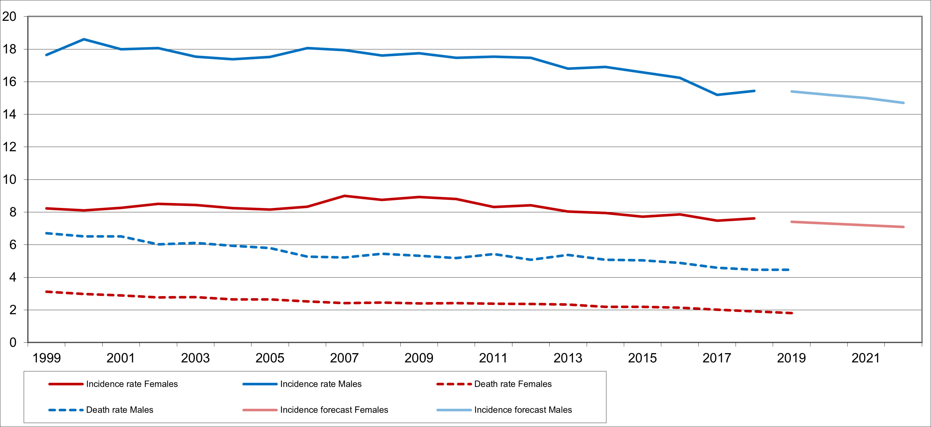
Renal cell carcinoma is one of the more common malignant tumors in adults. In Europe, men are affected significantly more often than women, with an incidence of approximately 26 per 100,000 compared to approximately 12 per 100,000. The median age at diagnosis is between 65 and 70 years for men and over 70 years for women. In recent years, renal cell carcinomas have been increasingly discovered incidentally during abdominal imaging for other indications, using ultrasound or cross-sectional imaging techniques. Since 2006, age-standardized incidence and mortality rates have been declining slightly.
The most effective treatment methods are Surgery—particularly in the localized stage—and drug therapy. Surgery with complete tumor resection is the only curative option. For systemic tumor therapy in metastatic disease, numerous new drugs in the fields of antiangiogenesis, tyrosine kinase inhibition, and immune checkpoint inhibition have been approved over the past 15 years, both as monotherapies and combination therapies. In palliative settings, particularly in cases of symptomatic, inoperable metastases, radiation therapy is also used.
2Basics
2.1Definition and Basic Information
Renal cell carcinoma accounts for approximately 85% of malignant kidney tumors. Other forms include urothelial carcinoma originating in the renal pelvis (10%), non-Hodgkin lymphomas, sarcomas, and, in children, nephroblastomas (Wilms tumor). This chapter focuses on renal cell carcinoma.
2.2Epidemiology
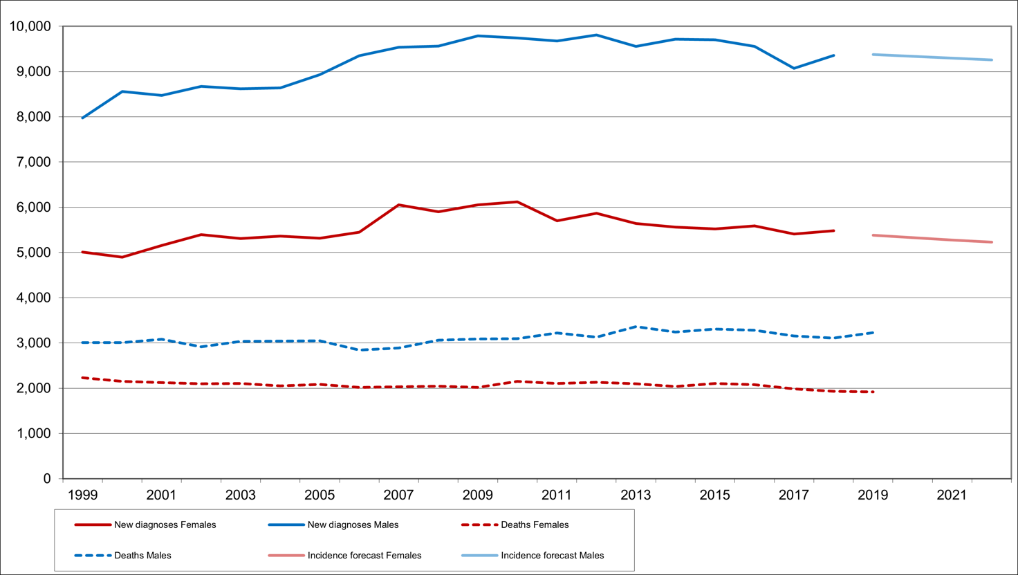
Renal cell carcinoma accounts for more than 99% of all malignant kidney tumors with specific histological characteristics. Sarcomas, nephroblastomas, and neuroendocrine tumors of the kidney are very rare. The following data therefore pertain to malignant kidney tumors (ICD-10: C64). Most recently (up to 2023), approximately 15,000 new cases of kidney cancer were diagnosed annually in Germany [1]; in Austria, the figure was 1,575 in 2024 [2]; and in Switzerland, there were approximately 1,200 cases per year most recently (2018–2022) [3]. Men are affected about twice as often as women. There are approximately 100,000 people living in Germany who have been diagnosed with kidney cancer in the past 10 years. Slightly fewer than 5,000 deaths per year in Germany are attributable to malignant kidney tumors; in Austria, the figure is around 400, and in Switzerland, around 300 cases [4]. Age-standardized incidence rates, as well as mortality rates, are declining for both sexes (Figure 1). Age-standardized mortality rates have also declined in Austria and Switzerland over the past few decades. The absolute 5-year survival rate is reported as 69% (men) and 68% (women); the relative 5-year survival rate, which takes into account mortality in the age-matched general population, is 80% (men) and 77% (women). The relative 10-year survival rate is 73% (men) and 70% (women) [1]. Approximately 60% of cases with sufficient staging information are diagnosed at Stage I; in these cases, the prognosis is very good. Even at Stage III, relative survival rates are still around 75% (Figure 2)
2.3Pathogenesis
Renal cell carcinoma is a heterogeneous disease. Histologically, clear cell, papillary, and chromophobe carcinomas are the most common [5]. The pathophysiology of renal cell carcinoma is characterized by the dysregulation of various signal transduction pathways. Clear cell carcinomas account for approximately 75–80% of tumors. They exhibit significant inter- and intratumoral heterogeneity. In approximately 80% of cases, functional inactivation of the von Hippel-Lindau (VHL) gene is present. This leads to activation of hypoxia-inducible factor (HIF)-1α and 2α, and increases the expression of genes involved in neoangiogenesis and cell proliferation. However, inactivation of the VHL gene alone is not sufficient for the development of renal cell carcinoma. Mutations are also found, albeit less frequently, in the PBRM1 (40%), SETD2 (15%), and BAP1 (15%) genes [6]. In a subgroup of clear-cell renal cell carcinomas, components of the mTOR (mechanistic/mammalian Target of Rapamycin) signaling pathway are altered at various levels. Furthermore, there are a multitude of epigenetic changes that have been shown in studies to have both prognostic and predictive value [7].
Papillary renal cell carcinomas (pRCC) are associated with alterations in the MET gene. The rare hereditary form is caused by a germline mutation in the MET oncogene on chromosome 7 [8].
In chromophobe renal cell carcinoma, aneuploidy with the loss of specific chromosomes is particularly common [9]. Mutations are frequently found in TP53, PTEN, FAAH2, PDHB, PDXDC1, and NZF765.
In the microenvironment, neoangiogenesis and the immune response offer targets for targeted therapies.
2.4Risk Factors
The risk of developing renal cell carcinoma is increased by the following factors:
Hereditary renal cell carcinomas account for approximately 5% of patients. To date, more than 12 genetically defined clinical syndromes have been identified. Germline mutations can be detected in 6–9% of newly diagnosed renal cell carcinomas [13]. The best-known syndromes are:
von Hippel–Lindau syndrome [OMIM 193300, autosomal dominant]: predisposition to clear cell renal cell carcinoma
Birt-Hogg-Dubé syndrome [OMIM 135150, autosomal dominant]: predisposition to chromophobe renal cell carcinoma
Acquired [14]
Obesity
Chronic kidney disease
Smoking
hypertension
Occupational exposure: halogenated hydrocarbons, long-term exposure to X-rays
3Prevention and Early Detection
3.1Prevention
The effectiveness of prevention is unclear. However, based on the underlying risk factors for renal cell carcinoma, general recommendations for prevention apply:
Do not smoke (nicotine abstinence)
Avoid being overweight
3.2Early Detection
There is no early detection program. Genetic counseling and an individualized monitoring strategy are recommended for family members of those with Hippel-Lindau syndrome as well as for young patients.
4Clinical characteristics
4.1Symptoms
Renal cell carcinoma is often asymptomatic. Locally related symptoms may include painless gross hematuria, flank pain, a palpable mass, or a newly developed varicocele. General signs of the disease include weight loss, fatigue, anemia, and paraneoplastic syndromes such as polycythemia, fever of unknown origin, neuropathy, or hypercalcemia. Many patients with renal cell carcinoma remain asymptomatic or have few symptoms for an extended period of time.
4.2Incidental Findings
In recent years, up to 50% of renal cell carcinomas have been discovered incidentally during abdominal imaging for other indications, using ultrasound or cross-sectional imaging. These asymptomatic tumors tend to be at an earlier stage [10]. Symptoms caused by metastases correspond to the sites of predilection: bone pain with skeletal involvement, cough and dyspnea with pulmonary involvement, and neurological deficits with cerebral or spinal involvement.
5Diagnosis
5.1Diagnostic Criteria
5.2Diagnosis
A thorough medical history and complete physical examination form the basis of rational diagnosis. The next step is to confirm the suspected clinical and/or imaging diagnosis; see Table 1.
If the suspected diagnosis of renal cell carcinoma has been confirmed by imaging, staging is indicated; see Table 2. Distant metastases can occur in almost any part of the body. The most common sites are the lungs, skeleton, liver, and brain.
A biopsy is indicated when it will influence the subsequent therapeutic approach, e.g., prior to local ablative procedures or prior to systemic therapy in cases of primary metastatic disease. Similarly, a biopsy may be indicated for assessing malignancy in small kidney tumors <2 cm—so-called “small renal masses”— , as the basis for a potential active surveillance strategy, particularly in older patients and those with comorbidities [15].
Furthermore, histological confirmation prior to surgical intervention is not required.
5.3Classification
5.3.1Histology
Histopathological classification follows the current WHO classification [5]; see Table 3.
Sarcomatous dedifferentiation can occur in all histological subgroups and should be documented. Other pathohistological classifications are prognostically relevant but have not yet influenced surgical strategy or the forms of drug therapy.
5.3.2Stages
5.4Prognostic Factors
5.4.1Prognosis score for metastatic renal cell carcinoma
Various models have been developed for the calculation and standardized assessment of risk factors. The so-called MSKCC or Motzer score has been validated in patients treated with chemotherapy and interferon [18, 19]; see Table 5.
In more recent studies, the IMDC score (International Metastatic Renal-Cell Carcinoma Database Consortium Score) is primarily used. It was developed during the TKI era and is based on the identification of 6 independent prognostic factors; see Table 6 [20].
Each risk factor is assigned one point; the IMDC score summarizes these [20].
The IMDC score is predictive of the choice of systemic therapy.
6Treatment
6.1Treatment structure
The most effective causal treatment methods are Surgery and drug therapy. Surgery is the only curative option. The overall treatment plan should be established before the first therapeutic measure is taken. A treatment algorithm is shown in Figure 3.
1 if surgically feasible;
2 minimally invasive, if possible;
3 in individual cases;
4 Indication depends on general condition, risk group, histology, and other factors;
5 no benefit in intermediate- and high-risk patients compared to sunitinib alone;
6 significant prolongation of disease-free survival (DFS) and overall survival (OS)
6.1.1Locally confined stages
The treatment of choice for locally confined renal cell carcinoma is surgical resection.
6.1.1.1Surgery
6.1.1.1.1Kidney
Radical and partial nephrectomy are available as alternatives. The previous gold standard was open radical nephrectomy with resection of Gerota’s fascia, the ipsilateral adrenal gland, and regional lymph nodes. The goal of partial nephrectomy is to preserve functional kidney tissue. Postoperative renal insufficiency is a negative prognostic factor [22].
In a randomized EORTC study of patients with clinically and radiologically suspected stage cT1/2 N0 renal cell carcinoma, the 10-year survival rate was 81.1% for those who underwent radical nephrectomy versus 75.7% for those who underwent partial nephrectomy. While a significant difference (p=0.03) was calculated in the intention-to-treat (ITT) analysis, it was not significant for patients with renal cell carcinoma after verification of the inclusion criteria (p=0.07). Based on these data, from Phase II studies with long-term follow-up, and a systematic review [23], the following recommendations can be derived:
Indications for partial nephrectomy [23]:
anatomical or functional solitary kidney
increased risk of renal insufficiency due to other causes (e.g., hypertension, diabetes mellitus)
hereditary renal cell carcinoma syndromes
Stage T1
In stage T2, the success of a partial nephrectomy depends on careful patient selection and surgical expertise.
Both radical and partial nephrectomy can be performed using open or minimally invasive techniques (retroperitoneoscopic, laparoscopic, or robot-assisted). Laparoscopic nephrectomy is less invasive and may reduce the risk of perioperative morbidity [24]. However, there is a lack of large randomized trials comparing the oncological equivalence of open and laparoscopic partial nephrectomy. Endoscopic procedures should be performed at select centers with appropriate expertise. Whenever oncologically feasible, kidney preservation via partial nephrectomy should be preferred over the radical procedure.
6.1.1.1.2Adrenal Gland
Adrenalectomy is only necessary if tumor infiltration or metastases are suspected based on imaging or intraoperative findings [24].
6.1.1.1.3Lymph Nodes
6.1.1.2Other local treatment modalities
6.1.1.2.1Embolization
Tumor embolization is used to reduce bleeding complications in the following situations:
as a standalone palliative measure in cases of persistent gross hematuria, when neither Surgery nor systemic therapy is possible due to poor general condition
in individual cases prior to surgical resection of locally advanced tumors
during the resection of bone metastases.
6.1.1.2.2Minimally invasive, ablative procedures
Various physical procedures are used for percutaneous, targeted therapy under imaging guidance [26, 27]. Tumor control rates of up to 85% after one year can be achieved through cryotherapy and radiofrequency ablation. Laser therapy and high-intensity focused ultrasound (HIFU) are less effective. Controlled comparative studies with long-term follow-up are lacking. These physical procedures are experimental. A prerequisite for their use is prior confirmation of the diagnosis by biopsy. Relative contraindications for local ablative procedures include a life expectancy of less than 1 year, multiple metastases, a low likelihood of success, tumors near the hilum, tumors larger than 5 cm, and tumors in the immediate vicinity of the calyces or the proximal ureter. Absolute contraindications include coagulation disorders or severe comorbidities.
6.1.1.3Adjuvant Therapy
Most studies in the adjuvant setting for various immunotherapy approaches, such as interferon or tumor vaccines, have yielded negative results. Several randomized trials with tyrosine kinase inhibitors (Assure, S-TRAC, PROTECT) showed no significant improvement in disease-free survival (DFS), with the exception of sunitinib in the S-TRAC trial [28, 29, 30]; a positive effect on overall survival has not yet been demonstrated.
In the adjuvant trial with pembrolizumab administered for 1 year in patients at high risk of recurrence (i.e., tumor stage 2 with Fuhrman grade 4 or sarcomatoid differentiation; tumor stage 3 or higher, regional lymph node metastases, or stage M1 with NED following metastasis resection) following nephrectomy, there was a significant prolongation of disease-free survival compared with placebo (HR 0.68 (0.53–0.87), p=0.002) [31]. At 24 months, the disease-free survival rate was 77.3% vs. 68.1%. The study was conducted only in patients with clear-cell renal cell carcinoma. Meanwhile, a benefit of adjuvant therapy with pembrolizumab is also emerging in terms of overall survival [32].
Adjuvant therapy with pembrolizumab should therefore be administered to patients with renal cell carcinoma who are at high risk of recurrence (i.e., stage 2 tumor with Fuhrman grade 4 or sarcomatoid differentiation; stage 3 or higher tumor, regional lymph node metastases, or stage M1 with NED following metastasis resection) [33].
Further Phase III studies of adjuvant therapy with checkpoint inhibitors such as atezolizumab [33] or nivolumab plus ipilimumab [34] showed no benefit in DFS and OS compared with placebo. The prospective randomized Litespark-22 trial comparing the combination of pembrolizumab and belzutifan versus pembrolizumab alone in patients at high risk of recurrence showed an increase in disease-free survival from 80.7% to 73.7% at 24 months, but no OS benefit [35]. Other studies are still ongoing.
6.1.2Locally Advanced Stages
One area of ongoing research is the treatment of patients with locally advanced carcinomas in whom complete resectability appears questionable based on imaging findings. The effectiveness of newer systemic therapies has led to approaches involving primary (neoadjuvant) systemic therapy followed by Surgery. These patients should be treated within the framework of clinical trials. To date, no benefit of neoadjuvant therapy has been demonstrated with respect to patient-relevant endpoints such as operability, progression-free survival, and overall survival. It also remains unclear which of the available agents should be preferred.
6.1.3Metastatic Renal Cell Carcinoma
The primary focus of treatment is systemic tumor therapy; see figure 4. A complementary cytoreductive nephrectomy may be discussed as part of a multimodal treatment strategy, depending on the risk of progression, during an interdisciplinary tumor board meeting; see chapter 6. 2. 1. 1. Cytoreductive Nephrectomy. Other local therapeutic procedures, such as radiation therapy for bone metastases or stereotactic radiation therapy, may be used as part of symptom-oriented measures; see chapter 6. 2. 3. Palliative Therapy—Symptom-Oriented.
6.1.3.1Drug Therapy
Treatment of metastatic renal cell carcinoma is almost always palliative. Before initiating drug therapy, the possibility of a watch-and-wait approach should be considered in patients with low or intermediate risk who are asymptomatic, particularly if there is no evidence of progression on follow-up imaging. With a watch-and-wait approach, regular clinical and imaging follow-ups at intervals of at least three months are recommended. Significant progress, with a marked prolongation of progression-free survival compared to the previous standard of care, interferon-alpha, has been achieved with angiogenesis-inhibiting multi-tyrosine kinase inhibitors (TKIs), mTOR inhibitors [35], the combination of interferon-alpha and the VEGF antibody bevacizumab, as well as, more recently, by newer TKIs and checkpoint inhibitors (ICI). Information on the use of these medications is summarized in the Appendix: approval status.
6.1.3.1.1First-line treatment of clear-cell renal cell carcinoma
The approaches to first-line drug therapy for locally advanced and metastatic renal cell carcinoma have changed fundamentally over the past two years. Various combination therapies and monotherapies are now available. It is important to note that most first-line studies included only clear-cell renal cell carcinoma (ccRCC) or renal cell carcinoma with a clear-cell component. In contrast, for non-clear-cell renal cell carcinoma (nccRCC), there are only a few studies with limited sample sizes, so the evidence is significantly weaker in this area. However, there are also studies in this area that primarily enrolled these patients, mostly in Phase II settings. These studies showed that, for the most part, the approaches used to treat clear-cell carcinoma are also effective for the other subtypes, albeit with slightly lower efficacy than for the clear-cell variant [36].
The efficacy of drug therapy, particularly with regard to overall survival, varies across the different risk groups according to the IMDC score. A treatment algorithm for drug therapy is shown in figure 4.
1 For risk scores, see chapter 5.4.1
2 Axitinib + avelumab: An OS benefit over sunitinib has only been demonstrated in the subgroup of high-risk patients
The majority of currently published randomized trials compare the respective new therapy with sunitinib monotherapy. Based on these data and the approval status, the combinations of nivolumab with cabozantinib [37], pembrolizumab with axitinib [38], pembrolizumab with lenvatinib [39], or—with certain restrictions—avelumab with axitinib [40] as the new standard of care in first-line therapy, regardless of risk factor or histological type, although there are insufficient data for non-clear-cell renal cell carcinoma and no improvement in OS data for the combination of axitinib with avelumab. For patients with intermediate- and high-risk disease, the combination of ipilimumab and nivolumab [41] represents an equivalent alternative. In the absence of comparative studies, it is currently not possible to establish a priority among these options. Data are summarized in table 7.
Study | Checkmate 214 [41] | Checkmate 9ER [37] | Keynote 426 [38] | CLEAR [39] | JAVELIN Renal 101 [40] |
|---|---|---|---|---|---|
Immunotherapy combination | Ipilimumab/Nivolumab | Nivolumab/Cabozantinib | Pembrolizumab/Axitinib | Pembrolizumab/Lenvatinib | Avelumab/ Axitinib |
Primary study endpoints | ORR, PFS, OS in patients with intermediate- and high-risk disease | PFS | OS and PFS in the ITT cohort | PFS | PFS and OS in patients with PD-L1-positive tumors (>1% of immune cells) |
ORR (%) | 39.0* | 55.7 | 59.3 | 71.0 | 51.4 |
CR (%) | 10.2* | 8.0 | 5.8 | 16.1 | 3.4 |
Primary Progress (%) | 20* | 5.6 | 5.4 | 11.5 | |
Median PFS (months) Immunotherapy combination vs. sunitinib |
12.4* vs. 12.3 P<0.001 |
16.6 vs. 8.3 P < 0.001 |
15.1 vs. 11.1 P<0.001 |
23.9 vs. 9.2 P<0.001 |
13.8 vs. 8.4 P < 0.0001 |
DoR (months) Combination immunotherapy vs. sunitinib | 76.2 vs. 25.1 P < 0.001 | 23.1 vs. 15.1 P=n.a. | 23.6 vs. 15.3 P=n.a. | 16.7 vs. 14.7 P=n.a. | - |
OS (months) Immunotherapy combination vs. sunitinib |
NR vs. 32.0 P<0.001 |
Median NR P = 0.001 |
Median NR P<0.001 |
Median NR P=0.005 |
Median NR n.s. |
The results for the various risk groups can be summarized as follows:
Low Risk of Progression
In the low-risk group, the combinations of nivolumab/cabozantinib, axitinib/pembrolizumab, and pembrolizumab/lenvatinib result in a significant increase in the remission rate and a prolongation of progression-free survival compared to sunitinib; however, a significant prolongation of overall survival compared to sunitinib has not yet been demonstrated.
Axitinib/Avelumab results in an increase in the remission rate and a prolongation of progression-free survival in the low- and intermediate-risk groups compared to sunitinib; however, a significant prolongation of overall survival compared to sunitinib has not yet been demonstrated.
Nivolumab + ipilimumab is inferior to sunitinib in terms of remission rate and progression-free survival (HR 2.18; median 9.8 months); the difference in overall survival is not significant.
Alternatives in cases of contraindications for these combinations are:Tyrosine kinase inhibitors: Sunitinib, pazopanib, and tivozanib are approved. The comparator arms in the respective approval studies varied.
Compared with interferon-alpha, sunitinib leads to an increase in the remission rate and a prolongation of progression-free survival (median 6 months).
In a non-inferiority study, pazopanib showed no significant difference in progression-free survival or overall survival compared to sunitinib, but had a slightly different side effect profile.
Compared with sorafenib, tivozanib results in a higher remission rate and longer progression-free survival (HR 0.795; median 2.4 months), but does not prolong overall survival.
Intermediate and high risk of progression
The combinations nivolumab/cabozantinib, axitinib/pembrolizumab, and pembrolizumab/lenvatinib resulted in a significant increase in the remission rate, as well as longer progression-free survival and overall survival, compared with sunitinib in the intermediate- and high-risk groups
In the intermediate-risk group, nivolumab plus ipilimumab resulted in an increase in the remission rate and a prolongation of overall survival compared with sunitinib (HR 0.697; median not yet reached); the difference in progression-free survival was not significant.
Axitinib + avelumab results in an increase in the remission rate and a prolongation of progression-free survival in low- and intermediate-risk patients compared to sunitinib, but not in overall survival (HR 0.87; median survival not yet reached).
Alternatives in cases of contraindications for these combinations are tyrosine kinase inhibitors:Sunitinib resulted in an increase in the remission rate and a prolongation of progression-free survival (median 6 months) compared with interferon-alpha.
In a small study, cabozantinib resulted in an increase in the remission rate and a prolongation of progression-free survival (HR 0.48; median 3.3 months) compared with sunitinib, but not in overall survival.
Details of the respective approval studies, including an assessment of clinical benefit according to the ESMO Magnitude of Clinical Benefit Scale (ESMO MCBS) and the G-BA’s previous benefit assessment, can be found in the fact sheets.
6.1.3.1.2First-line therapy for non-clear-cell renal cell carcinoma
According to the EU approval, non-clear-cell renal cell carcinomas can be treated with TKI/ICI combinations in randomized trials—similar to clear-cell renal cell carcinomas—despite limited data. Entity-specific treatment recommendations cannot currently be provided.
In a small prospective randomized trial comparing sunitinib with cabozantinib, crizotinib, and savolitinib, cabozantinib significantly prolonged progression-free survival (PFS) compared with sunitinib (9.0 vs. 5.6 months), although no benefit in overall survival (OS) was observed [42].
There are several single-arm studies evaluating a TKI/IO combination. A Phase II study of cabozantinib plus nivolumab reported an objective response rate (ORR) of 47.5%, with a median progression-free survival (PFS) of 12.5 months and a median overall survival (OS) of 28 months [43]. The single-arm NEMESIS trial evaluated pembrolizumab plus axitinib as first-line therapy and demonstrated a disease control rate of 78.1% and an ORR of nearly 44% in patients with pRCC and chromophobic RCC; the median PFS was 10.8 months, while the median OS has not yet been reached [44]. In the Phase II KEYNOTE-B61 study, which evaluated pembrolizumab plus lenvatinib as first-line therapy for various nccRCC subtypes, 93 of 158 patients had pRCC. The ORR in this subgroup was 54% (9% complete remissions (CR) and 45% partial remissions (PR)) with a median PFS of 17.5 months. The 12-month OS rate for the entire nccRCC population was 63%; the median OS has not yet been reached [45].
In the prospective randomized trial (Sunniforecast) comparing ipilimumab and nivolumab versus “standard of care” (SOC) in 309 patients with non-clear-cell renal cell carcinoma, a significant improvement in the 12-month survival rate was demonstrated (78% vs. 68%; p=0.026), along with a nominal improvement in median survival time (33.2 vs. 25.2 months). An exploratory analysis showed, in particular, a survival benefit for patients with a CPS-PDL1 score >1 (HR = 0.56; p = 0.008) [46]. This randomized trial thus represents the highest level of evidence to date. In the papillary subgroup, which was confirmed by reference pathology, the 12-month OS rate was 74.8% in the ipilimumab/nivolumab arm versus 63.4% in the SOC arm, and the median OS was 24.9 months versus 18.9 months [47].
6.1.3.1.3Second-line therapy
Due to the introduction of combination therapies in the first-line setting and the lack of controlled studies in the second-line setting following combination therapy, an evidence-based recommendation cannot be provided. Second-line therapy should therefore be determined based on individual assessment (e.g., prior therapy, response, disease course, comorbidities).
Following first-line therapy with immune checkpoint inhibitors and their combination with a TKI or another immune checkpoint inhibitor, there are currently no evidence-based data regarding the subsequent treatment sequence. Agents not used in first-line therapy may be tried in second-line and subsequent lines of treatment.
In patients initially treated with a TKI, nivolumab results in a higher remission rate and longer progression-free survival (HR 0.40; median 4.6 vs. 4.2 months), prolongs overall survival (HR 0.51; median 25.5 vs. 19.6 months), and reduces the rate of severe adverse events (CTCAE Grade 3/4) [48].
Cabozantinib also leads to an increase in the remission rate, a prolongation of progression-free survival (HR 0.58; median 7.4 vs. 3.8 months), and a prolongation of overall survival (HR 0.7; median 21.4 vs. 17.1 months). The rate of severe CTCAE Grade 3/4 adverse events is higher [49, 50].
In a small study of patients primarily treated with a TKI, lenvatinib plus everolimus, compared with everolimus alone, resulted in an increase in the remission rate, prolonged progression-free survival (HR 0.4; median 14.6 vs. 5.5 months), and prolonged overall survival (HR 0.51; median 25.5 vs. 15.4 months). The rate of severe adverse effects (CTCAE Grade 3/4) is higher. Data from a follow-up study using a lower lenvatinib dose (14 mg vs. 18 mg) show similar toxicity but a trend favoring the higher dose in terms of ORR, PFS, and OS [51, 52].
Agents not used in first-line therapy may be tried in second-line and subsequent lines of treatment. It can therefore be assumed that drugs effective in first-line treatment or following VEGF-targeted therapy will retain their efficacy even after the new combinations. Prospective studies or, at the very least, registry data are urgently needed in this regard. A randomized study comparing tivozanib with sorafenib in patients who had previously been treated with VEGFR and immune checkpoint inhibitors showed a slight prolongation of progression-free survival (HR 0.73; median 1.7 months), but not of overall survival. Currently, treatment recommendations are primarily based on the type of prior treatment, the patient’s general condition, and the side effects of previous therapies; see Figure 4.
Depending on the treatment goal, comorbidities, and side effects of previous therapies, other TKIs and the mTOR inhibitor everolimus may also be used.
A newer agent is belzutifan, an oral, selective HIF-2α inhibitor. Belzutifan is approved for patients with familial renal cell carcinoma due to a von Hippel-Lindau (VHL) gene mutation and for patients with advanced clear-cell renal cell carcinoma following prior therapy with a PD-1 or PD-L1 inhibitor and a VEGF TKI [53, 54].
Details of the respective approval studies, including an assessment of clinical benefit according to the ESMO Magnitude of Clinical Benefit Scale (ESMO MCBS) and the G-BA’s previous benefit assessment, can be found in the fact sheets.
6.2Treatment modalities
6.2.1Surgical Treatment Options
6.2.1.1Cytoreductive nephrectomy
In patients with advanced renal cell carcinoma, nephrectomy may lead to regression of metastases; however, this phenomenon has been observed in fewer than 2% of patients. When combined with systemic therapy using interferon-alpha, nephrectomy prolongs median survival by 3 to 10 months.
In a non-inferiority study of metastatic patients with intermediate- and high-risk disease, sunitinib alone was not inferior to cytoreductive nephrectomy followed by sunitinib; in fact, there was a trend toward improved overall survival (OS) with sunitinib alone [55]. The value of sequential tumor nephrectomy was also investigated in the SURTIME study. The study did not meet its primary endpoint; a total of 99 patients were randomized [56]. By selecting patients who responded to TKI therapy, the additional surgery could be avoided in patients with an unfavorable prognosis (progression within 4 months).
The results of modern treatment strategies using TKIs or immune checkpoint inhibitors were primarily achieved in patients who had undergone nephrectomy. To date, no data are available on the value and sequence of tumor nephrectomy in combination with immune checkpoint inhibitors and combination therapies (ICI/TKI or ICI/ICI).
6.2.1.2Resection of Metastases
Long-lasting remissions have been observed following resection of metastases, particularly in the lungs, liver, and brain. Therefore, this procedure is recommended—following careful staging—for patients in whom an R0 resection is possible [28, 33, 57, 58, 59]. The decision to proceed with surgical therapy must be made on an individual basis and must take into account factors such as comorbidities, prognosis, and the patient’s wishes. Follow-up imaging to detect any new metastases should be performed prior to metastasis surgery in order to assess the disease progression and the appropriateness of metastasis resection. Conceptually, surgical metastasis resection should be performed with the goal of complete resection of the tumor burden or solely for palliative purposes. Debulking surgery should be used only for symptom control or in cases of impending or manifest complications. Even after systemic therapy, a subsequent complete metastasis resection can still achieve a long-term treatment-free interval.
In cases of initial complete resection of the primary tumor and the metastases (“no evidence of disease,” NED), adjuvant therapy with pembrolizumab is indicated (see chapter 6.1.1.3 above).
6.2.1.3Radiation Therapy for Metastases
Renal cell carcinoma is not very sensitive to radiation. No randomized studies are available. Nevertheless, indications for CyberKnife or stereotactic radiation therapy may arise in cases of solitary or oligometastases. This applies primarily to brain metastases, or in individual cases to metastases in other organs [28, 60, 61, 62, 63].
Current data show that even inoperable localized renal cell carcinoma can be successfully treated with stereotactic radiation therapy, achieving durable local control with low toxicity. However, randomized studies are also lacking in this area to further evaluate its efficacy [1].
6.2.2Drug Therapy (in alphabetical order)
6.2.2.1Avelumab
Avelumab is a human IgG1 monoclonal antibody. It binds to programmed cell death ligand 1 (PD-L1) and prevents its binding to its receptor, PD-1. A PD-1/PD-L1 receptor-ligand interaction leads to the inhibition of CD8+ T cells and thus to the suppression of the immune response. Avelumab is approved in combination with axitinib for first-line treatment of metastatic renal cell carcinoma. Compared with sunitinib, this combination results in a higher response rate (51.4% vs. 25.7%) and prolonged progression-free survival (13.8 vs. 8.4 months; HR 0.69). Side effects of avelumab monotherapy are relatively rare. In monotherapy for Merkel cell carcinoma, severe side effects classified as CDTAE Grade 3 or 4 were limited exclusively to laboratory values; see Avelumab. The most common side effects of all grades were fatigue (24%), infusion reactions (17%), diarrhea (9%), asthenia (8%), rash (7%), and loss of appetite (6%). Possible immune-mediated reactions occurred at Grade 1/2: hypothyroidism (3%), hyperthyroidism (2%), pneumonitis (1%), and type 1 diabetes mellitus (1%). The side effects of combination therapy are consistent with those of axitinib and other immune checkpoint inhibitors (see also under nivolumab).
6.2.2.2Axitinib
Axitinib is a second-generation tyrosine kinase inhibitor. It selectively blocks VEGF receptors 1–3. In second-line therapy, remission rates of 19% and a significantly longer progression-free survival time compared to the control group were achieved. Overall survival was not prolonged. Severe adverse effects (Grade 3/4) occurring in more than 5% of patients included hypertension (16%), diarrhea (11%), and fatigue (11%). Patients treated with multikinase inhibitors over the long term may experience endocrine (hypothyroidism), hematologic, or cardiac adverse effects.
6.2.2.3Belzutifan
Belzutifan is an inhibitor of hypoxia-inducible factor 2-alpha (HIF-2α). It is an oral “small molecule” drug. In patients with renal cell carcinoma associated with VHL syndrome, an overall response rate of 49% (CI 36% to 62%) was observed. In pretreated patients who had received a PD-1 or PDL-1 inhibitor and a VEGF-TKI, the overall response rate was significantly improved compared with everolimus (21.9% vs. 3.5%), as were the PFS rates at 12 and 18 months (33.7% vs. 17.6% and 22.5% vs. 9.0%, respectively). OS did not differ significantly.
6.2.2.4Cabozantinib
Cabozantinib is a multikinase inhibitor. In addition to the VEGFR1, VEGFR2, and VEGFR3 kinases, it also inhibits AXL and MET. Cabozantinib is approved for advanced renal cell carcinoma as first-line monotherapy (does not apply to Switzerland) for patients with intermediate- and high-risk disease and as second-line therapy at a dose of 60 mg/day. In the registration trial, following prior VEGFR-targeted therapy, cabozantinib resulted in longer overall survival (HR 0.67; median 4.9 months) and progression-free survival (HR 0.52; median 3.5 months) compared with everolimus, as well as a higher remission rate. The rate of severe treatment-related adverse events is significantly higher with cabozantinib than with everolimus; CTCAE Grade 3/4 adverse events that occurred more frequently than in the everolimus arm were hypertension (15%) and fatigue (9%). The most common adverse events leading to dose reduction with cabozantinib were diarrhea (16%), palmar-plantar erythrodysesthesia (11%), and fatigue (10%). In the registration trial, 60% of patients treated with cabozantinib required a dose reduction.
6.2.2.5Everolimus
Everolimus is an oral mTOR inhibitor. The registration study was conducted in patients receiving second-line or later-line therapy following prior treatment with sorafenib and/or sunitinib and demonstrated a significant prolongation of progression-free survival compared with the placebo control group. Two-thirds of the patients had also been pretreated with cytokines. Severe adverse events (Grade 3/4) that occurred in more than 5% of patients in the registration trial were infections (10%) and dyspnea (7%). A less common but debilitating adverse effect of mTOR inhibitors is pneumonitis.
6.2.2.6Ipilimumab
Ipilimumab is a humanized monoclonal antibody that targets the CTLA-4 protein. Its use can reverse the negative immune regulation caused by CTLA-4 and achieve an antitumor effect through T-cell stimulation. For renal cell carcinoma, ipilimumab was tested in combination with nivolumab in a Phase III trial, based on studies in other tumors, particularly melanoma. Compared with sunitinib, the combination demonstrated an increased response rate (42% vs. 27%) in intermediate- and high-risk patients, as well as prolonged progression-free survival (HR 0.83) and overall survival (HR 0.63). In patients with a low risk of progression, the nivolumab/ipilimumab combination was inferior to sunitinib. Grade 3/4 CTCAE adverse events occurring in more than 1% of patients in the nivolumab/ipilimumab arm included fatigue (4%), elevated lipase (10%), and diarrhea (4%). Treatment was discontinued in 22% of patients in the nivolumab/ipilimumab arm due to adverse events.
6.2.2.7Lenvatinib
Lenvatinib is a multikinase inhibitor that inhibits the VEGFR1, VEGFR2, and VEGFR3 kinases. Lenvatinib is approved for first-line treatment of advanced renal cell carcinoma in combination with pembrolizumab at a dose of 20 mg/day orally, in combination with pembrolizumab 200 mg intravenously every 21 days. Compared with sunitinib, this combination results in a significant increase in overall response rate (71.0% vs. 36.1%), progression-free survival (23.9 months vs. 9.2 months; HR 0.39 (0.32–0.49); p<0.001) and overall survival (HR 0.66 (0.49–0.88); p=0.005). The rate of severe Grade 3/4 adverse events was slightly higher with the combination (82.4% vs. 71.8%), with hypertension (27.6%), diarrhea (9.7%), and weight loss (8.0%) being the most common.
In second-line treatment, lenvatinib is approved as a combination therapy with everolimus at a dose of 18 mg/day plus everolimus at a dose of 5 mg/day. The previous benefit assessment was based on a three-arm Phase IIb/II study involving a total of 153 patients. Compared with everolimus alone, lenvatinib/everolimus in second-line therapy resulted in prolonged overall survival (HR 0.51; median 10.1 months), progression-free survival (HR 0.40; median 9.1 months), and the remission rate. The rate of severe treatment-related adverse events was significantly higher with lenvatinib/everolimus than with everolimus. Grade 3/4 adverse events that occurred more frequently than in the everolimus arm included diarrhea (20%), fatigue (14%), hypertension (14%), vomiting (8%), nausea (6%), proteinuria (4%), and back pain (4%).
6.2.2.8Nivolumab
Nivolumab is a monoclonal anti-PD-1 antibody. Nivolumab blocks apoptosis in activated T cells and enhances the autologous immune response. Nivolumab is approved for first-line therapy in combination with ipilimumab. The combination of nivolumab and ipilimumab demonstrates an increased response rate (42% vs. 27%) compared to sunitinib in patients with intermediate and high risk of progression, as well as prolonged progression-free survival (HR 0.83) and overall survival (HR 0.63). Grade 3/4 CTCAE adverse events occurring in more than 1% of patients in the nivolumab/ipilimumab arm included fatigue (4%), elevated lipase (10%), and diarrhea (4%). Treatment was discontinued in the nivolumab/ipilimumab arm in 22% of patients due to adverse events.
Nivolumab is approved as monotherapy for second-line treatment of metastatic renal cell carcinoma. In second-line therapy, nivolumab results in prolonged survival (HR 0.73; median 5.4 months), an increased remission rate, and a longer time to clinical symptom progression compared with everolimus. Progression-free survival is not significantly prolonged. The rate of severe treatment-related adverse events is significantly lower with nivolumab than with everolimus, and the rate of treatment discontinuations is also lower. Grade 3/4 CTCAE adverse events with nivolumab included fatigue (2%), anemia (2%), diarrhea (1%), dyspnea (1%), pneumonitis (1%), and hyperglycemia (1%). Fatigue (33%), nausea (14%), pruritus (14%), diarrhea (12%), loss of appetite (12%), and rash/acne (10%) were also the most common side effects overall with nivolumab.
6.2.2.9Pazopanib
Pazopanib is another oral tyrosine kinase inhibitor with a slightly different kinase profile than sorafenib and sunitinib. The registration trial included both patients receiving first-line therapy and those who had previously been treated with cytokines. The response rate was 30%, and progression-free survival was significantly longer than in the placebo control group. Overall survival was not prolonged. There were no severe adverse effects (Grade 3/4) occurring in more than 5% of patients in the registration trial. Regular monitoring of ALT and bilirubin is recommended for the early detection of hepatic toxicity. Patients treated with multikinase inhibitors over the long term may experience endocrine (hypothyroidism), hematologic, or cardiac side effects.
6.2.2.10Pembrolizumab
Pembrolizumab is a humanized IgG4 monoclonal antibody. It binds to the programmed cell death receptor (PD-1) and prevents the binding of its ligands, such as PD-L1. A PD-1/PD-L1 receptor-ligand interaction leads to the inhibition of CD8+ T cells and thus to the suppression of the immune response; pembrolizumab counteracts this negative regulation. Pembrolizumab is approved in combination with axitinib for first-line treatment of metastatic renal cell carcinoma. Compared with sunitinib, this combination results in a higher response rate (59.3% vs. 35.7%), prolonged progression-free survival (15.1 vs. 11.1 months; HR 0.69), and prolonged overall survival (HR 0.53; median not yet reached). The side effects are similar to those of other immune checkpoint inhibitors (see under Nivolumab).
6.2.2.11Sorafenib
Sorafenib is an oral inhibitor of several tyrosine kinases, including VEGF receptors, PDGFRB, Flt-3, and c-KIT. In signal transduction, it also blocks serine-threonine kinases of the Raf family in the MAPK pathway. In the largest study to date with sorafenib, this agent was evaluated as a second-line therapy in patients with low or intermediate risk. Progression-free survival was significantly prolonged. In first-line therapy, there was no significant difference in remission rate or progression-free survival compared with interferon-alpha. A severe adverse effect (Grade 3/4) that occurred in more than 5% of patients in the registration trial was hand-foot syndrome (Grade 3/4). Patients treated with multikinase inhibitors over the long term may experience endocrine (hypothyroidism), hematologic, or cardiac adverse effects.
6.2.2.12Sunitinib
Sunitinib is an oral inhibitor that blocks several VEGF and PDGF receptors, as well as c-KIT and Flt-3, at the tyrosine kinase level. In the registration trial, sunitinib was used in patients receiving first-line therapy in comparison with IFN-alpha. Progression-free survival was significantly longer, and the remission rate was 47% in the final analysis. Severe adverse effects (Grade 3/4) that occurred in more than 5% of patients in the registration trial included hypertension (12%), fatigue (11%), diarrhea (11%), hand-foot syndrome (9%), and asthenia (7%). In patients treated with multikinase inhibitors over the long term, endocrine (hypothyroidism), hematologic, or cardiac side effects may occur.
6.2.2.13Temsirolimus
Temsirolimus was the first approved mTOR kinase inhibitor for renal cell carcinoma. The drug is administered intravenously. Its efficacy was evaluated in a randomized Phase III trial in patients with at least 3 of 6 risk factors (Table 5). Patients in the comparator arm were treated with IFN-alpha, while patients in a third arm received temsirolimus plus IFN-alpha. Treatment with temsirolimus resulted in remission rates of 8.6%; median progression-free survival and overall survival were significantly prolonged compared with monotherapy with IFN-alpha. The combination showed no benefit over temsirolimus monotherapy; however, the dose of temsirolimus was reduced to 15 mg per week in the combination arm. Severe adverse events (Grade 3/4) occurring in more than 5% of patients in the registration trials included anemia (20%), asthenia (11%), hyperglycemia (11%), and dyspnea (9%). A less common but debilitating side effect of mTOR kinase inhibitors is pneumonitis.
6.2.2.14Tivozanib
Tivozanib is another oral tyrosine kinase inhibitor that selectively inhibits VEGF receptors. In the pivotal trial, tivozanib was compared with sorafenib and resulted in prolonged progression-free survival in first-line therapy (12.7 vs. 9.1 months) and overall survival (11.9 vs. 9.1 months) (hazard ratio 0.756 for first-line therapy, p=0.037). The remission rate increased to 33.1% compared with 23.4%. Tivozanib did not prolong overall survival; however, the data are of limited interpretability due to a 61% switching (crossover) rate from the sorafenib arm to the tivozanib arm. Grade 3/4 adverse events that occurred in ≥5% of patients receiving tivozanib in the registration trial were hypertension (27%), fatigue (5%), and elevated lipase levels (9%). Another common adverse event is dysphonia.
6.2.2.15Cytostatic Agents
Conventional cytostatic agents have only limited efficacy in renal cell carcinoma. Agents used included 5-fluorouracil in combination with immunotherapy or vinblastine. Remission rates with chemotherapy were below 5%.
6.2.3Sequential therapy, new options
The new drug treatment options for metastatic renal cell carcinoma have profoundly changed the clinical picture of the disease and the management of patients. For the majority of patients, several drugs with different mechanisms of action will be used in sequence as the disease progresses. The optimal sequence has not yet been established. The choice of medications should therefore be guided by the treatment goal and the patient’s overall clinical condition and comorbidities, taking into account the expected treatment-related side effects.
6.3Special Situations
6.3.1Non-clear-cell renal cell carcinoma
Clear-cell renal cell carcinoma is the dominant histological entity. The majority of studies with newer drugs have been conducted exclusively in this subgroup. Patients with type II papillary renal cell carcinoma have a more aggressive course of disease and a shorter life expectancy. Analyses of this subgroup suggest that they respond to kinase inhibitors and antiangiogenic therapy, albeit with lower remission rates and shorter progression-free survival.
It is recommended that patients with non-clear-cell renal cell carcinoma be treated according to the algorithm for clear-cell carcinomas. This also applies to the use of immune checkpoint inhibitors.
If possible, treatment within the framework of clinical trials should be considered. For these patients, a short-term evaluation is indicated to allow for a change in the mechanism of action in the event of non-response.
6.3.2Palliative Care—Symptom-Oriented
Palliative care involves the individualized, symptom-oriented treatment of physical and psychological symptoms at every stage of the disease course. It is provided through an interdisciplinary approach, and in particular, the inclusion of psycho-oncological care should be considered. The necessity and options for palliative care should be discussed early and thoroughly with all those involved. The following specific symptoms occur particularly frequently in patients with advanced renal cell carcinoma.
6.3.2.1Bone Metastases
In addition to adequate and tailored pain management, local and systemic measures are available for the treatment of patients with bone metastases. In cases of a single bone metastasis, surgical treatment with a primarily curative intent should be performed. In cases of pain or risk of fracture, radiation therapy is the treatment of choice. It can be administered in a hypofractionated regimen alongside ongoing systemic therapy. An additional option is surgical intervention for pathological fractures, unstable vertebral fractures, or to relieve spinal compression.
Systemic measures include causal therapy and the administration of bone-modifying agents (bisphosphonates, anti-RANKL antibodies). They reduce the risk of complications and delay the progression of bone metastasis. There are no prospective randomized trials conducted exclusively in patients with renal cell carcinoma or in a sufficiently large cohort. Information on the approval status of bone-modifying agents can be found in the appendix “Approval for Renal Cell Carcinoma (in German only).” Bisphosphonates are also indicated for hypercalcemia.
6.3.2.2Liver and Lung Metastases
The focus is on causal, systemic therapy. In individual cases, local therapy may be indicated. In addition to surgical resection, local ablative procedures are available. Prerequisites are:
no disseminated metastases
no local recurrence or clinically limiting secondary cancer
Decisions regarding the local treatment of liver or lung metastases are made by interdisciplinary tumor boards.
6.3.2.3Brain metastases
The first-line treatment for symptomatic metastases is the administration of steroids to reduce perifocal edema. For isolated, resectable brain metastases, local surgical therapy is recommended. An alternative is targeted local conformal radiation therapy (stereotactic radiation therapy, Gamma Knife, CyberKnife). Partial or whole-brain radiation therapy may be considered for patients with disseminated brain metastases who are in good general health, have no extracerebral disease progression, and have a life expectancy of more than 3 months—preferably with preservation of the hippocampus to avoid cognitive toxicity. Data on the efficacy of newer drugs are limited to small patient cohorts. In the non-randomized CABRAMET study with cabozantinib, partial remission was reported in 16 of 26 (61.5%) patients with brain metastases; median survival was 15.8 months [64].
7Rehabilitation
All patients should be offered specialty-specific rehabilitation in the form of follow-up treatment (AHB) or follow-up rehabilitation (ARH, AR) after therapy for renal cell carcinoma. In cases of persistent or newly occurring treatment-related complications, patients should be informed about additional rehabilitation options. Patients with metastatic disease also benefit from specialized rehabilitation. The goals of medical rehabilitation are to maintain or restore the ability to work, lead an independent daily life, prevent the need for long-term care, and participate in social life. Rehabilitation should be multidisciplinary and utilize multimodal treatment approaches, tailored to the patient’s comorbidities. As part of the rehabilitation program, patients should be offered targeted physical therapy, psycho-oncological care to support coping with the disease, and social-medical counseling; in cases of functional limitations, occupational therapy should also be provided. When recommending a rehabilitation clinic, the patient’s wishes (right to request and choose a facility under Section 9 of SGB IX) should be taken into account, with consideration given to the clinic’s oncological focus.
8Follow-up Care and Follow-up
8.1Follow-up
Follow-up care after primary tumor therapy in the non-metastatic stage should be risk-adapted [33]. In the first year, a follow-up every 3 months (clinical examination, laboratory tests, and ultrasound) is recommended; in the second year, a follow-up every 6 months; and from years 3–5, an annual follow-up is recommended. Particularly after partial nephrectomy, ultrasound assessment is complex and difficult; therefore, risk-adapted cross-sectional imaging (abdominal CT) is recommended for these patients; see also the recommendations of the S3 guideline [33].
During ongoing systemic therapy, imaging should be performed every 6 to 12 weeks. Treatment with checkpoint inhibitors may initially lead to an increase in tumor mass, known as early pseudoprogression. For this reason, the first imaging study in these patients is often indicated only 12 weeks after the start of therapy.
8.2Postoperative Follow-Up for Patients with Locally Advanced Renal Cell Carcinoma
There is no universally applicable follow-up program. The risk of relapse depends on the stage at initial diagnosis. The majority of relapses occur within the first two years. Since life expectancy in the event of relapse is influenced by the extent of metastasis, follow-up with cross-sectional imaging appears reasonable. However, there is no evidence that structured follow-up in the form of regular staging examinations leads to improved survival. The goal of examinations following curative therapy is to detect complications and long-term effects. In patients who have undergone nephrectomy, these primarily include symptoms of renal insufficiency and hypertension.
9References
https://www.krebsdaten.de/krebs/de/content/publikationen/krebs_in_deutschland/kid_2025/kid_2025_c64_niere
https://www.statistik.at/statistiken/bevoelkerung-und-soziales/gesundheit/krebserkrankungen
https://nkrs.ch/de/statistiken-und-berichte/statistiken-zu-krebs
https://ec.europa.eu/eurostat/databrowser/view/hlth_cd_aro/default/table?lang=en&category=hlth.hlth_cdeath.hlth_cd_gmor
Moch H, Amin MB, Berney DM et al. The 2022 World Health Organization classification of tumors of the urinary system and male genital organs - Part A: Renal, penile, and testicular tumors. Eur Urol 2022;82:458-468. DOI:10.1016/j.eururo.2022.06.016
The Cancer Genome Atlas Research Network. Comprehensive molecular characterization of clear cell renal cell carcinoma. Nature 2013;499:43-49. DOI:10.1038/nature12222
Joosten SC, Smits KM, Aarts MJ et al. Epigenetics in renal cell cancer: mechanisms and clinical applications. Nat Rev Urol 2018;15:430-451. DOI:10.1038/s41585-018-0023-z
The Cancer Genome Atlas Research Network. Comprehensive molecular characterization of papillary renal cell carcinoma. N Engl J Med 2016;374:135-145. DOI:10.1056/NEJMoa1505917
Ciccarese C, Brunelli M, Montironi R et al. The prospect of precision therapy for renal cell carcinoma. Cancer Treat Rev 2016;49:37-44. DOI:10.1016/j.ctrv.2016.07.003
Carlo MI, Hakimi AA, Stewart GD et al. Familial kidney cancer: implications of new syndromes and molecular insights. Eur Urol 2019;76:754-764. DOI:10.1016/j.eururo.2019.06.015
Shuch B, Vourganti S, Ricketts CJ et al. Defining early-onset kidney cancer: implications for germline and somatic mutation testing and clinical management. J Clin Oncol 2014;32:431–437. DOI:10.1200/JCO.2013.50.8192
Huang KL, Mashl RJ, Wu Y et al. Pathogenic germline variants in 10,389 adult cancers. Cell 2018;173: 355–370. DOI:10.1016/j.cell.2018.03.039
Carlo MI, Mukherjee S, Mandelker D et al. Prevalence of germline mutations in cancer susceptibility genes in patients with advanced renal cell carcinoma. JAMA Oncol 2018;4:1228–1235. DOI:10.1001/jamaoncol.2018.1986
Capitanio U, Bensalah K, Bex A et al. Epidemiology of renal cell carcinoma. Eur Urol 2019;75:74-84. DOI:10.1016/j.eururo.2018.08.036
Cheung DC, Finelli A. Active surveillance of small renal masses in the elderly: a literature review. Eur Urol Focus 2017;3:340-351. DOI:10.1016/j.euf.2017.11.005
Amin MB, Edge SB, Greene F et al (eds.). AJCC Cancer Staging Manual, 8th ed. New York: Springer International Publishing; 2017. ISBN 978-3-319-40617-6
Cohen HT, McGovern FJ. Renal-cell carcinoma. N Engl J Med 2005;353:2477-2490. DOI:10.1056/NEJMra043172
Motzer RJ, Bacik J, Murphy BA et al. Interferon-alpha as a comparator in clinical trials of new therapies for advanced renal cell carcinoma. J Clin Oncol 2002;20:289-296. DOI:10.1200/JCO.20.1.289
Heng DYC, Xie W, Regan MM et al. External validation and comparison with other models of the International Metastatic Renal-Cell Carcinoma Database Consortium prognostic model: a population-based study. Lancet Oncol 2013;14:141-148. DOI:10.1016/S1470-2045(12)70559-4
https://www.mdcalc.com/imdc-international-metastatic-rcc-database-consortium-risk-model-metastatic-renal-cell-carcinoma
Weight CJ, Larson BT, Fergany AF et al. Nephrectomy-induced chronic renal insufficiency is associated with an increased risk of cardiovascular death and death from any cause in patients with localized cT1b renal masses. J Urol 2010;183:1317-1323. DOI:10.1016/j.juro.2009.12.030
MacLennan S, Imamura M, Lapitan MC et al. Systematic review of oncological outcomes following surgical management of localized renal cancer. Eur Urol 2012;61:972-993. DOI:10.1016/j.eururo.2012.02.039
Smith ZL. Current status of minimally invasive surgery for renal cell carcinoma. Curr Urol Rep 2016;17:43. DOI:10.1007/s11934-016-0599-x
Bekema HJ, MacLennan S, Imamura M et al. Systematic review of adrenalectomy and lymph node dissection in locally advanced renal cell carcinoma. Eur Urol 2013;64: 799-810. DOI:10.1016/j.eururo.2013.04.033
Blom JHM, van Poppel H, Maréchal JM et al. Radical nephrectomy with and without lymph node dissection: final results of the European Organization for Research and Treatment of Cancer (EORTC) randomized phase 3 trial 30881. Eur Urol 2009;55:28–34. DOI:10.1016/j.eururo.2008.09.052
Ljungberg B, Albiges L, Abu-Ghanem Y et al. European Association of Urology guidelines on renal cell carcinoma: the 2022 update. Eur Urol 2022;82:399-410. DOI:10.1016/j.eururo.2022.03.006
S3 Guideline on the Diagnosis, Treatment, and Follow-up of Renal Cell Carcinoma. Long Version 5.0 – September 2024, AWMF Registration Number: 043-017OL. https://register.awmf.org/de/leitlinien/detail/043-017OL
Haas NB, Manola J, Uzzo RG et al. Adjuvant sunitinib or sorafenib for high-risk, non-metastatic renal cell carcinoma (ECOG-ACRIN E2805): a double-blind, placebo-controlled, randomized, phase 3 trial. Lancet 2016;387:2008-2016. DOI:10.1016/S0140-6736(16)00559-6
Ravaud A, Motzer RJ, Pandha HS et al. Adjuvant sunitinib in high-risk renal-cell carcinoma after nephrectomy. N Engl J Med 2015;375:2246-2254. DOI:10.1056/NEJMoa1611406
Motzer RJ, Haas NB, Donskov F et al. Randomized phase III trial of adjuvant pazopanib versus placebo after nephrectomy in patients with locally advanced renal cell carcinoma. J Clin Oncol 2017;35:3916-3923. DOI:10.1200/JCO.2017.73.5324
Choueiri TK, Tomczak P, Park SH et al. Adjuvant pembrolizumab after nephrectomy in renal cell carcinoma. N Engl J Med 2021;385:683-694. DOI:10.1056/NEJMoa2106391
Choueiri TK, Tomczak P, Park SH et al.; KEYNOTE-564 investigators. Overall survival with adjuvant pembrolizumab in renal-cell carcinoma. N Engl J Med 2024;390:1359-1371. DOI:10.1056/NEJMoa2312695
Pal SK, Uzzo R, Karam JA et al. Adjuvant atezolizumab versus placebo for patients with renal cell carcinoma at increased risk of recurrence following resection (Immotion010): a multicenter, randomized, double-blind, phase 3 trial. Lancet 2022;400:1103-1116. DOI:10.1016/S0140-6736(22)01658-0
Motzer RJ, Russo P, Grünwald V et al. Adjuvant nivolumab plus ipilimumab versus placebo for localized renal cell carcinoma after nephrectomy (CheckMate 914): a double-blind, randomized, phase 3 trial. Lancet 2023;401:821-832. DOI:10.1016/S0140-6736(22)02574-0
Choueiri TK, Motzer RJ, Karam JA et al.; LITESPARK-022 Investigators. Adjuvant pembrolizumab plus belzutifan for renal-cell carcinoma. N Engl J Med 2026;395:32-43
Bergmann L, Weber S, Hartmann A, Ahrens M. Pathology and systemic therapy of non-clear cell renal cell carcinoma: an overview. Expert Rev Anticancer Ther 2021;21:1273-1286. DOI:10.1080/14737140.2021.1959319
Choueiri TK, Powles T, Burotto M et al. Nivolumab plus cabozantinib versus sunitinib for advanced renal-cell carcinoma. N Engl J Med 2021;384:829-841. DOI:10.1056/NEJMoa2026982
Rini BI, Plimack ER, Stus V et al. Pembrolizumab plus axitinib versus sunitinib for advanced renal-cell carcinoma. N Engl J Med 2019;380:1116-1127. DOI:10.1056/NEJMoa1816714
Motzer R, Alekseev B, Rha SY et al. Lenvatinib plus pembrolizumab or everolimus for advanced renal cell carcinoma. N Engl J Med 2021;384:1289-1300. DOI:10.1056/NEJMoa2035716
Motzer RJ, Penkov K, Haanen J et al. Avelumab plus axitinib versus sunitinib for advanced renal-cell carcinoma. N Engl J Med 2019;380:1103-1115. DOI:10.1056/NEJMoa1816047
Motzer RJ, Tannir NM, McDermott DF et al. Nivolumab plus ipilimumab versus sunitinib in advanced renal-cell carcinoma. N Engl J Med 2018;378:1277-1290. DOI:10.1056/NEJMoa1712126
Barata P, Tangen C, Plets M et al. Final overall survival analysis of S1500: a randomized, phase II study comparing sunitinib with cabozantinib, crizotinib, and savolitinib in advanced papillary renal cell carcinoma. J Clin Oncol 2024;42:3911-3916. DOI:10.1200/JCO.24.00767
Fitzgerald KN, Lee CH, Voss MH et al. Cabozantinib plus nivolumab in patients with non-clear cell renal cell carcinoma: updated results from a phase 2 trial. Eur Urol 2024;86:90-94. DOI:10.1016/j.eururo.2024.04.025
Stellato M, Buti S, Maruzzo M et al. Pembrolizumab plus axitinib for metastatic papillary and chromophobe renal cell carcinoma: NEMESIA (non-clear metastatic renal cell carcinoma pembrolizumab axitinib) study, a subgroup analysis of the I-RARE observational study (Meet-URO 23a). Int J Mol Sci 2023;24:1096. DOI:10.3390/ijms24021096
Albiges L, Gurney H, Atduev V et al. Pembrolizumab plus lenvatinib as first-line therapy for advanced non-clear-cell renal cell carcinoma (KEYNOTE-B61): a single-arm, multicenter, phase 2 trial. Lancet Oncol 2023;24:881-891. DOI:0.1016/S1470-2045(23)00276-0
Bergmann L, Albiges L, Ahrens M et al. Prospective randomized phase II trial of ipilimumab/nivolumab versus standard of care in non-clear-cell renal cell carcinoma—results of the SUNNIFORECAST trial. Ann Oncol 2025;36:796-806. DOI:10.1016/j.annonc.2025.03.016
Ahrens M, Haanen J, Albiges L et al. Papillary renal cell carcinoma—exploratory results of the SUNNIFORECAST trial comparing ipilimumab plus nivolumab versus standard of care as first-line therapy based on central pathological review. Eur J Cancer 2026;243:116816. DOI:10.1016/j.ejca.2026.116816
Motzer RJ, Escudier B, McDermott DF et al. Nivolumab versus everolimus in advanced renal-cell carcinoma. N Engl J Med 2015;373:1803-1813. DOI:10.1056/NEJMoa1510665
Choueiri TK, Escudier B, Powles T et al. Cabozantinib versus everolimus in advanced renal-cell carcinoma. N Engl J Med 2015;373:1814-1823. DOI:10.1056/NEJMoa1510016
Motzer RJ, Escudier B, Powles T et al. Long-term follow-up of overall survival for cabozantinib versus everolimus in advanced renal cell carcinoma. Br J Cancer 2018;118:1176-1178. DOI:10.1038/s41416-018-0061-6
Motzer RJ, Hutson TE, Glen H et al. Lenvatinib, everolimus, and the combination in patients with metastatic renal cell carcinoma: a randomized, phase 2, open-label, multicenter trial. Lancet Oncol 2015;16:1473-1482. DOI:10.1016/S1470-2045(15)00290-9
Pal SK, Puente J, Heng DYC et al. Assessing the safety and efficacy of two starting doses of lenvatinib plus everolimus in patients with renal cell carcinoma: a randomized phase 2 trial. Eur Urol 2022;82:283-292. DOI:10.1016/j.eururo.2021.12.024
Jonasch E, Donskov F, Iliopoulos O et al. Belzutifan for renal cell carcinoma in von Hippel-Lindau disease. N Engl J Med 2021;385:2036-2046. DOI:10.1056/NEJMoa2103425,
Choueiri TK, Powles T, Peltola K et al.; LITESPARK-005 investigators. Belzutifan versus everolimus for advanced renal-cell carcinoma. N Engl J Med 2024;391:710-721. DOI:10.1056/NEJMoa2313906
Méjean A, Ravaud A, Thezenas S et al. Sunitinib alone or after nephrectomy in metastatic renal-cell carcinoma. N Engl J Med 2018;379:417-442. DOI:10.1056/NEJMoa1803675
Bex A, Mulders P, Jewett M et al. Comparison of immediate vs. deferred cytoreductive nephrectomy in patients with synchronous metastatic renal cell carcinoma receiving sunitinib: the SURTIME randomized clinical trial. JAMA Oncol 2019;5:164-170. DOI:10.1001/jamaoncol.2018.5543
Blanco-Fernández G, Fondevila-Campo C, Sanjuanbenito A et al. Pancreatic metastases from renal cell carcinoma. Postoperative outcome after surgical treatment in a Spanish multicenter study (PANMEKID). Eur J Surg Oncol 2022;48:133-141. DOI:10.1016/j.ejso.2021.08.011
Meacci E, Nachira D, Zanfrini E et al. Prognostic factors affecting survival after pulmonary resection of metastatic renal cell carcinoma: a multicenter experience. Cancers (Basel) 2021;13:3258. DOI:10.3390/cancers13133258
Internò V, De Santis P, Stucci LS et al. Prognostic factors and current treatment strategies for renal cell carcinoma that has metastasized to the brain: an overview. Cancers (Basel) 2021;13:2114. DOI:10.3390/cancers13092114
Alongi F, Arcangeli S, Triggiani L et al. Stereotactic ablative radiation therapy in renal cell carcinoma: from oligometastatic to localized disease. Crit Rev Oncol Hematol 2017;117:48-56. DOI:10.1016/j.critrevonc.2017.07.004
Miccio JA, Oladeru OT, Jun Ma S, Johung KL. Radiation therapy for patients with advanced renal cell carcinoma. Urol Clin North Am 2020;47:399-411. DOI:10.1016/j.ucl.2020.04.011
Yim K, Leeman JE. Radiation therapy in the treatment of localized and advanced renal cancer. Urol Clin North Am 2023;50:325-334. DOI:10.1016/j.ucl.2023.01.008
Siva S, Ali M, Correa RJM et al. 5-year outcomes after stereotactic ablative body radiotherapy for primary renal cell carcinoma: an individual patient data meta-analysis from IROCK (the International Radiosurgery Consortium of the Kidney). Lancet Oncol 2022;23:1508-1516. DOI:10.1016/S1470-2045(22)00656-8
Negrier S, Mourey L, Dalban C et al. Cabozantinib in patients with non-locally pretreated brain metastases from renal cell carcinoma: Results of the multicenter phase II trial CABRAMET. Eur J Cancer 2026;239:116712. DOI:10.1016/j.ejca.2026.116712
10[Kapitel nicht relevant]
11[Kapitel nicht relevant]
12[Kapitel nicht relevant]
13[Kapitel nicht relevant]
14Links
16Disclosure of Potential Conflicts of Interest
in accordance with the rules of the relevant professional societies.
Download
Reference:
Quellenangabe:
Onkopedia-Leitlinien werden kontinuierlich an den Stand des Wissens angepasst. Die jeweils gültige Version, AGB und Nutzungsbedingungen finden Sie unter www.onkopedia.com.
Für die kommerzielle Nutzung wenden Sie sich bitte an onkopedia@dgho.de.
Onkopedia guidelines are continuously adapted to the state of knowledge. The currently valid version, terms of use and general terms and conditions can be found at onkopedia-guidelines.info.
For commercial use, please contact onkopedia@dgho.de.